It combines scraping and heat.
A lesion is scraped with a curette and treated electrically.
Electrodesiccation and Curettage: A small, low-risk skin cancer is scraped out with a sharp loop, and the base is then burned. It takes one visit and needs no stitches. It leaves a round pale scar, and the edges are never checked under a microscope.
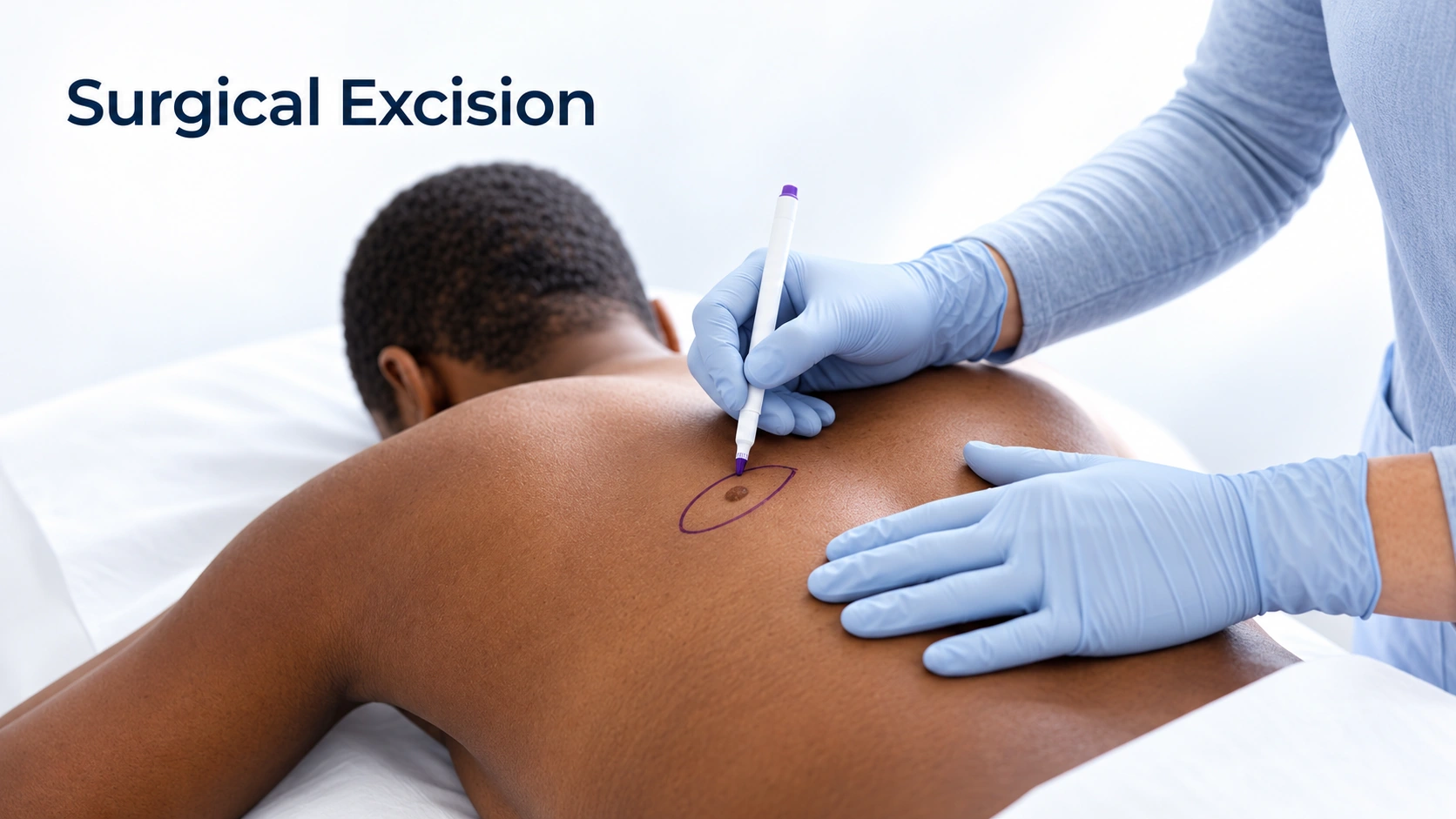
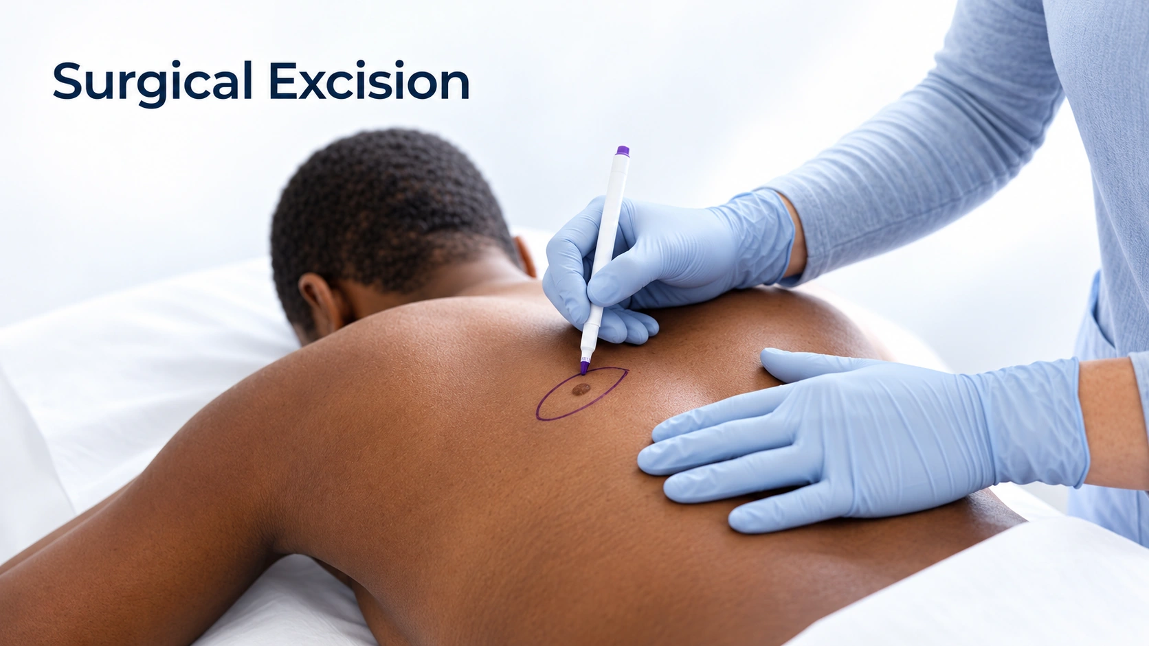
Surgical Excision: The growth is cut out along with a measured border of normal-looking skin, and the wound is stitched closed. The whole piece goes to a laboratory and the result comes back in about a week. The scar is a line, and it is longer than the spot was.

Choose for a small, low-risk skin cancer on the body when one quick visit with no stitches matters more than margin checking.
Choose to remove a skin cancer or suspicious growth completely with lab-checked edges — the scar is a line, longer than the spot.
Can you have both?
Both remove skin cancers. ED&C is quicker for small, low-risk cancers; excision gives stitches and a margin check.
A lesion is scraped with a curette and treated electrically.
It can treat carefully selected low-risk skin cancers.
It lacks the margin assessment of excision or Mohs surgery.
Type, site, size and depth determine suitability.
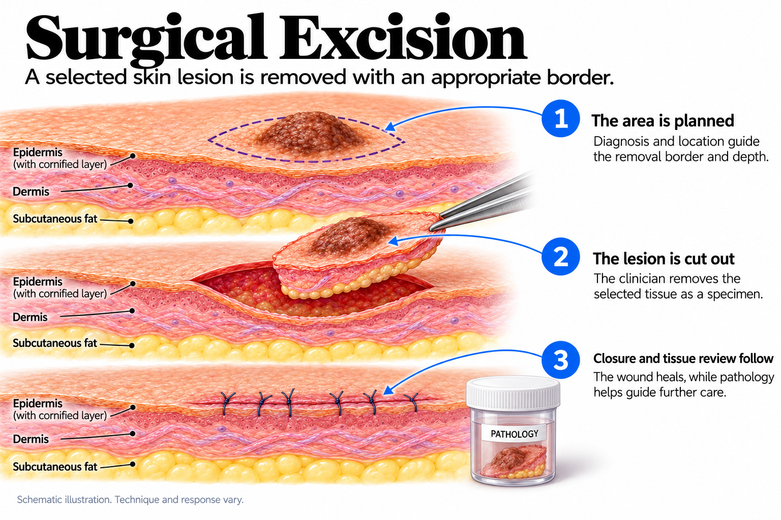
A planned incision removes the lesion and a rim of tissue.
It provides tissue or removes a known lesion.
They should not be chosen from a generic chart.
Standard excision and Mohs assess tissue differently.
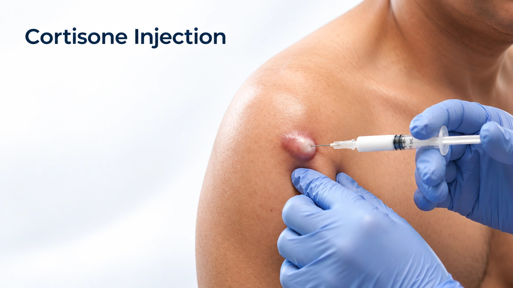
High cure rates for small tumors.
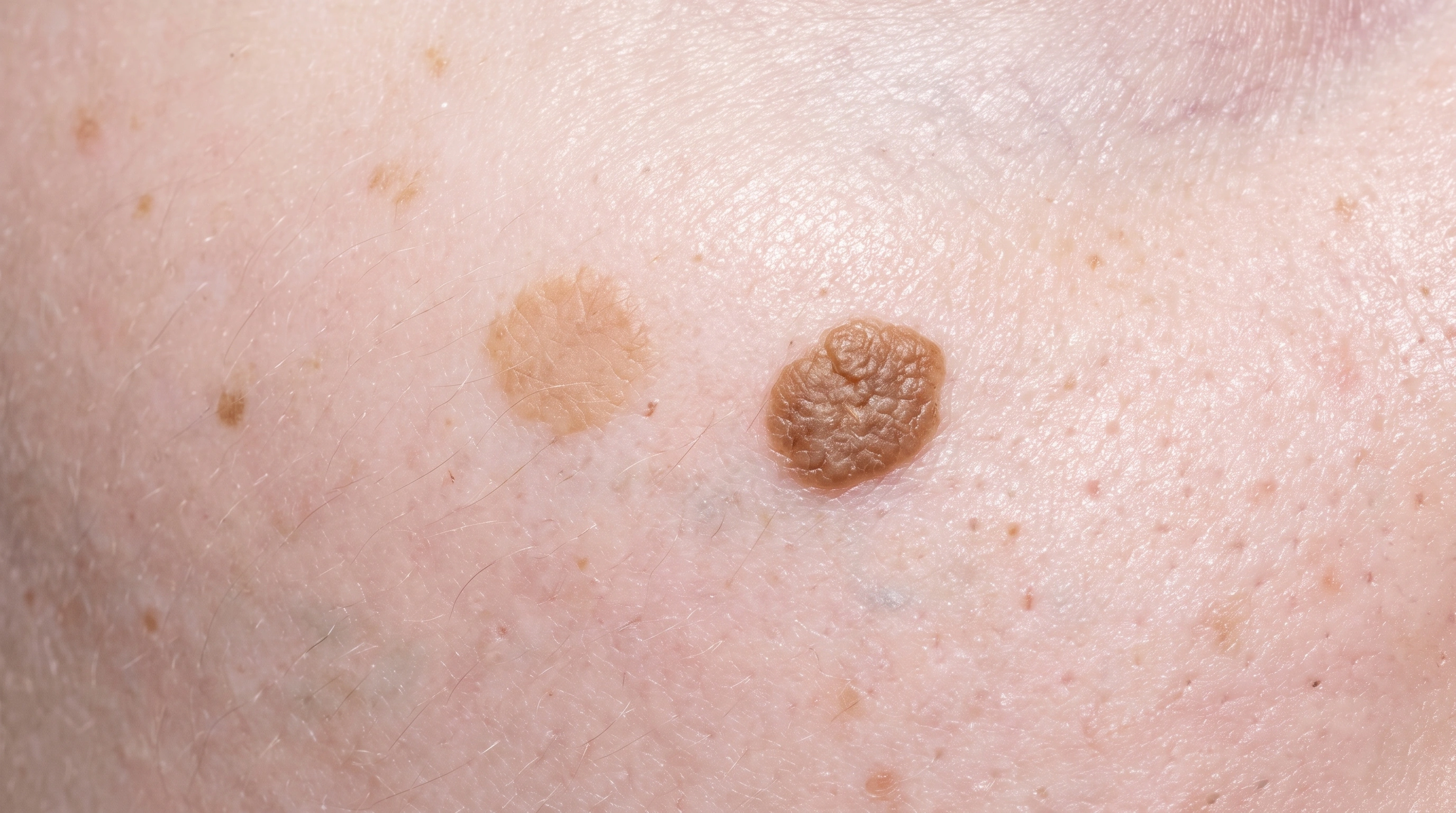
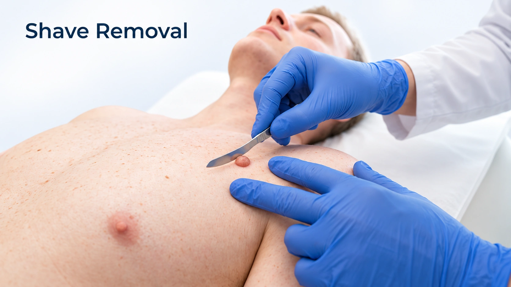
Removes growths quickly.
High cure rates.
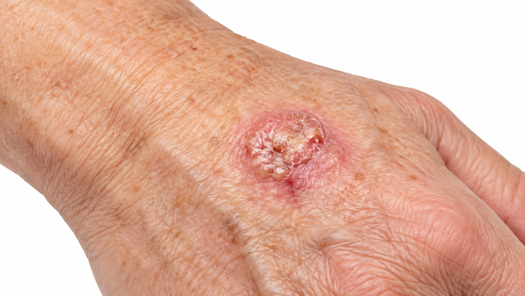
Removes the cancer with margins.
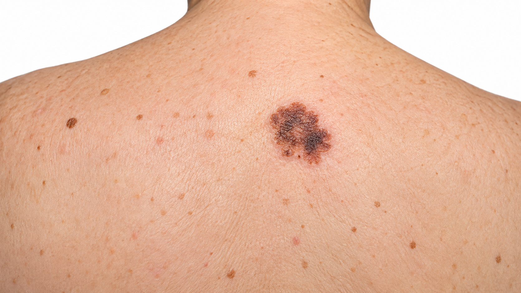
The main treatment.
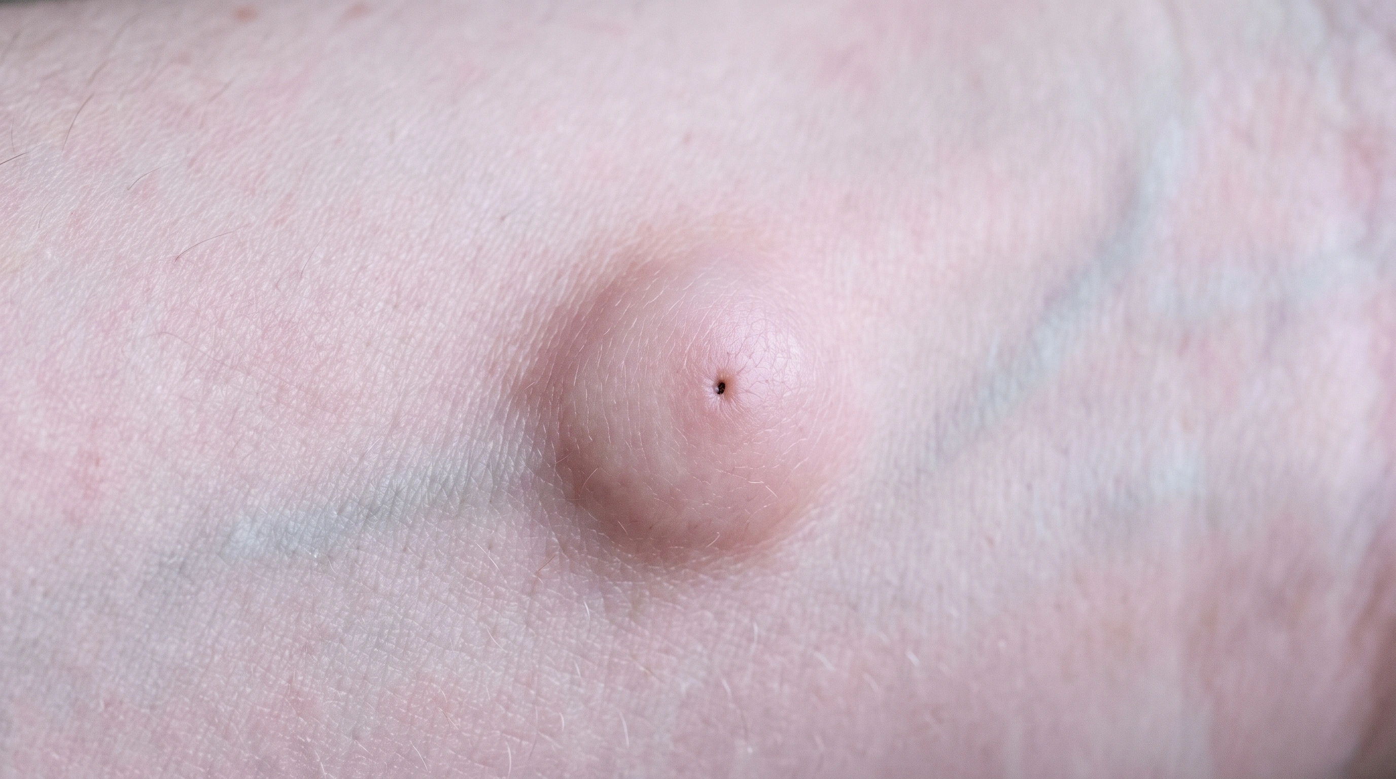
Removes the sac.
For small, low-risk tumors.
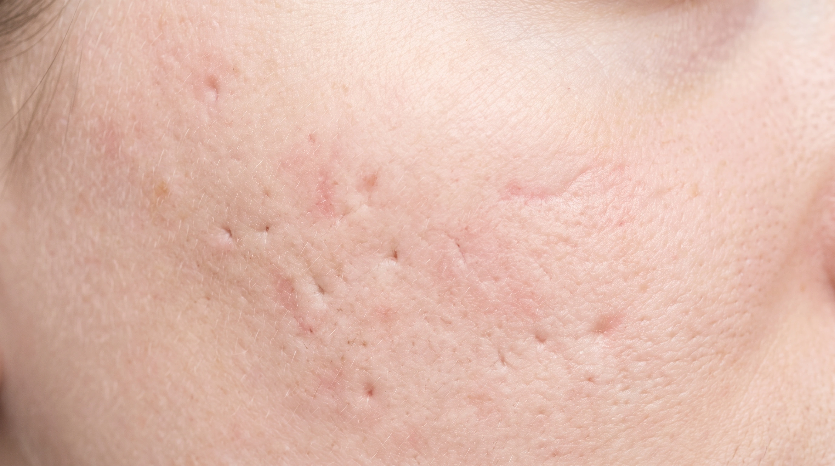
Punch excision is used for small scars instead.
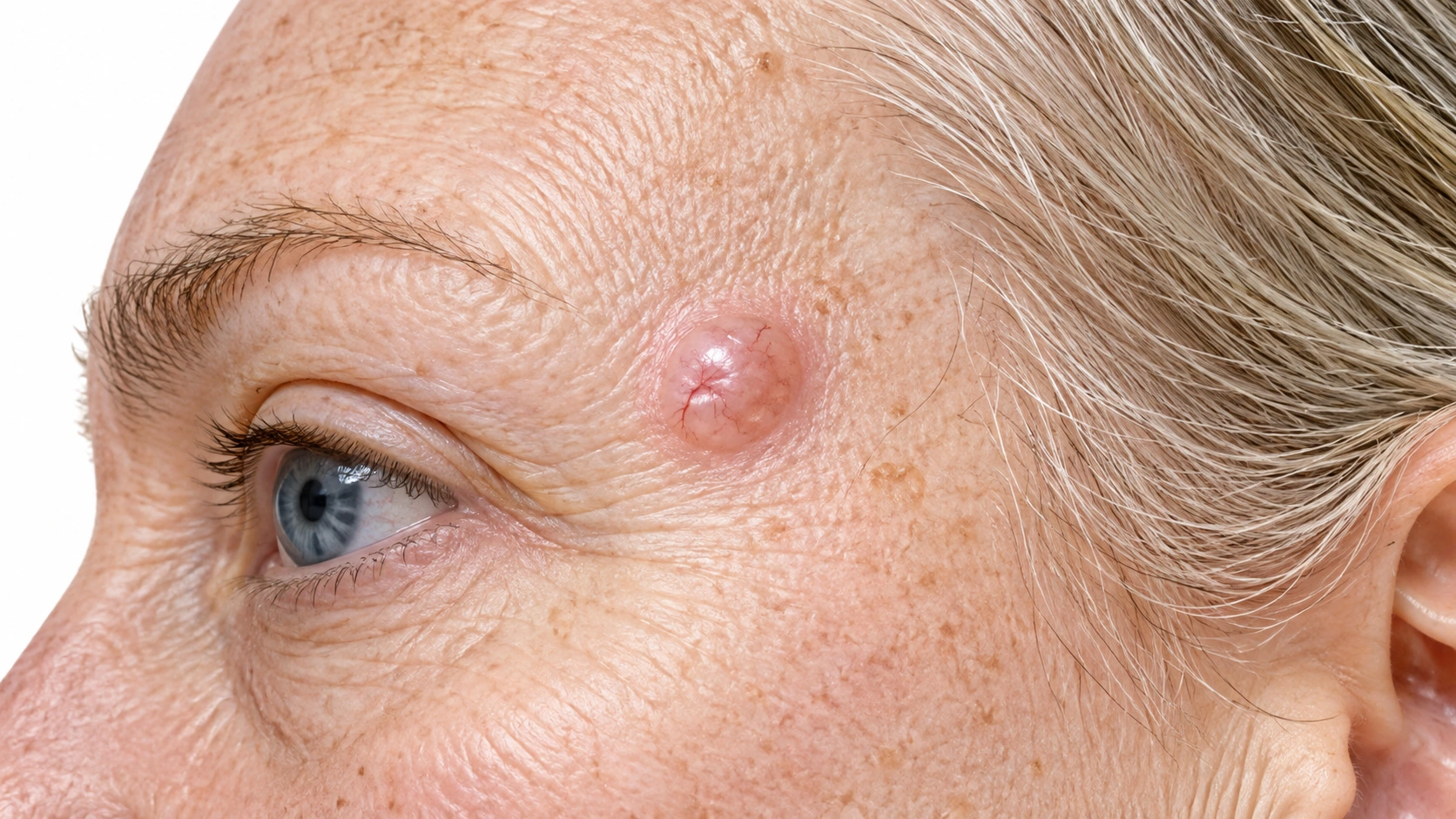
Needs excision with margins.
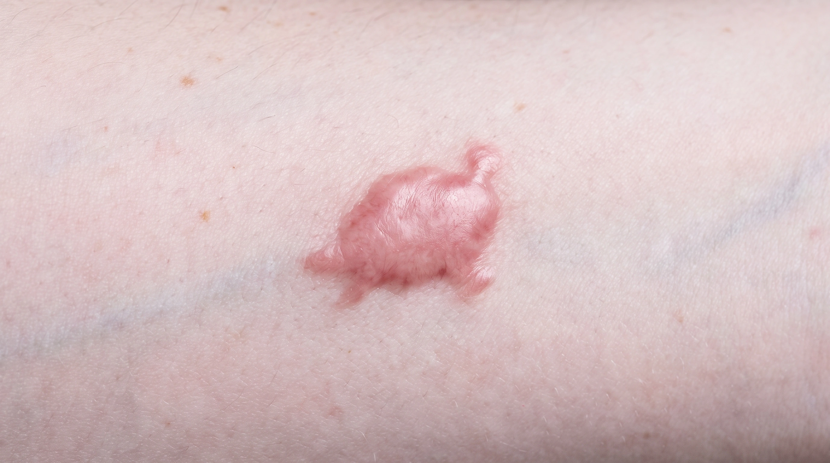
Can trigger new keloids.
Keloids often return larger alone.
The specific lesion, scar or affected patch determines the treatment site; there is no fixed body distribution.
The specific lesion, scar or affected patch determines the treatment site; there is no fixed body distribution.

A curette removes tissue: A clinician scrapes away the selected lesion. Electrical treatment treats the base: Energy dries or coagulates tissue in the treatment bed. The wound heals: The open area repairs with scar tissue; follow-up remains important.
Schematic illustration. Technique and response vary. This explains a mechanism and does not specify treatment settings.
The area is planned: Diagnosis and location guide the removal border and depth. The lesion is cut out: The clinician removes the selected tissue as a specimen. Closure and tissue review follow: The wound heals, while pathology helps guide further care.
Schematic illustration. Technique and response vary. This explains a mechanism and does not specify treatment settings.
Do not stop: Blood thinners: Aspirin, clopidogrel, warfarin, apixaban, rivaroxaban and similar are usually continued — extra oozing is easier to manage than a clot or a stroke.
Ointment and dressings: Buy plain petrolatum ointment and non-stick dressings beforehand, not on the way home.
Supplements and alcohol: Non-essential blood thinners such as fish oil, vitamin E and ginkgo are commonly stopped about a week before, and alcohol the night before.
Smoking and vaping: Stop for a week before and two weeks after if you can.
What to wear and eat: Wear something you do not mind getting blood on.
Eating and clothes: Eat normally and take your usual morning medicines.
The lesion is matched against the biopsy report, photographed, and consent signed.
The site is confirmed against the biopsy report and you point to it yourself.
The doctor scrapes with the curette, which feels like pressure and tugging, not pain.
The electric probe follows, with a faint buzz and a smell of burning. The pair is repeated two or three times; one lesion takes about 10 to 20 minutes. Nothing should feel sharp once you are numb, so say so if it does.
The surgery: The skin is cleaned and draped.
The surgery: The ellipse is cut out in one piece, bleeding points sealed, and the edges freed so they meet without tension. Closing is in layers — dissolving stitches deep, stitches on the surface. You feel pressure and tugging and hear instruments, but nothing sharp. Most take 20 to 45 minutes, and the site feels tight afterward rather than sore.
A round, shallow, raw crater slightly larger than the growth, under ointment and a dressing.
A dressing, written wound care, a stitch removal appointment, and a plan for how the pathology result reaches you.
Day 1 to 7: The wound weeps and the base looks yellow.
First week: Keep the dressing dry and in place for 24 to 48 hours, then start daily care — gentle wash, petrolatum, non-stick dressing.
Days 5 to 14: Stitches come out — face at 5 to 7 days, trunk and arms at 10 to 14, legs and back sometimes longer.
Weeks 2 to 6: The crater shrinks and the base turns pink.
From week 2: The scar is hardest, pinkest and most raised at around three to six weeks.
Month 2 to 12: The scar starts pink and firm, then flattens and pales over roughly a year.
Immediately
Lab result in about a week
90–95% cure for suitable cancers
Permanent
This is the least expensive way to treat a skin cancer surgically.
An excision costs more than a scrape-and-burn and less than Mohs surgery.
In the US it is billed as a destruction procedure, priced by lesion size and body site, and generally covered when it treats a diagnosed skin cancer.
In the US the surgical charge is priced by the size of the excision and the body site, with a separate charge for a more complex layered closure.
The numbing injection stings for under a minute. After that you feel pressure and tugging, not pain. Afterward the site is sore rather than painful, and acetaminophen is enough.
There is no intact specimen to check — the tumor is scraped away in fragments and the base burned. The method relies on the operator feeling soft tumor against firm normal tissue, which is reliable for shallow, clearly bordered tumors and unreliable for others.
A round, flat disc, usually slightly wider than the original spot and lighter than the skin around it. Some are a little sunken, some raised. Pink at first, paling over about a year. It is permanent.
About 3 to 6 weeks on the face, trunk and arms. On the lower leg it is slower, sometimes two to three months, particularly in older adults or with diabetes or poor circulation.
Sometimes, for a small superficial tumor on the cheek or forehead. Generally not for the nose, eyelids, ears, lips or the creases around them, where recurrence is higher and there is less spare skin. Ask why one is recommended over the other for your tumor.
For a well-selected, small, low-risk tumor, cure at 5 years is commonly quoted around 95 percent. That falls when the method is used on tumors it was not meant for — aggressive subtypes, recurrent tumors, unclear borders, high-risk facial sites.
Because a round hole cannot be closed flat. The piece taken out is an ellipse roughly three times as long as it is wide, so the edges meet evenly instead of puckering at each end. A spot the size of a pencil eraser becomes a line an inch or more long. It is geometry, not technique, and no surgeon avoids it.
Usually 3 to 10 days, depending on the laboratory. Ask before you leave how and when you will be told, and call if you have heard nothing after about two weeks.
A further procedure — a re-excision with a wider border, or a referral for Mohs surgery — usually within a few weeks. It is not a mistake. It is the reason the specimen went to a laboratory in the first place.
About 2 weeks for most sites, longer for the back, shoulder and shin. The visible stitches come out sooner, and the deep stitches are still carrying the tension. Lifting or straining is what stretches or splits a wound.
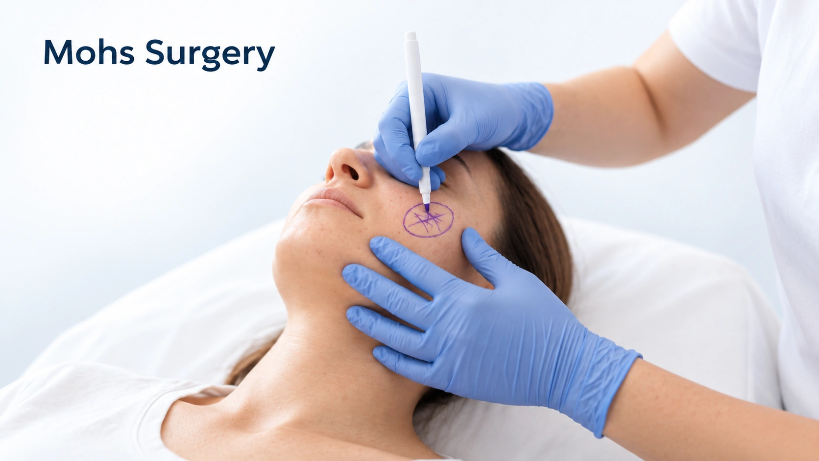
On the face, particularly the nose, eyelids, ears and lips, or for a tumor that is large, has vague borders, has an aggressive subtype on the biopsy, or has come back before, Mohs is often better. For a small low-risk cancer on the trunk or limbs, an excision achieves the same result in a fraction of the time and cost. Ask which category yours is in.
It flattens and pales substantially over about a year, with most of the change in the first six months. Keeping it out of the sun and using silicone gel or sheeting once it is closed are the two things with reasonable evidence behind them. Scars on the chest, shoulders and upper back stay red and raised longest and are the likeliest to widen.