
Melanoma vs Atypical Moles

Melanoma is a skin cancer that starts in the cells that make pigment. It is much less common than other skin cancers but causes most skin cancer deaths, because it can spread. Found and removed while it is still thin, it is usually curable. Benign moles with unusual clinical features that can resemble melanoma.

Research updated 2026-10-23
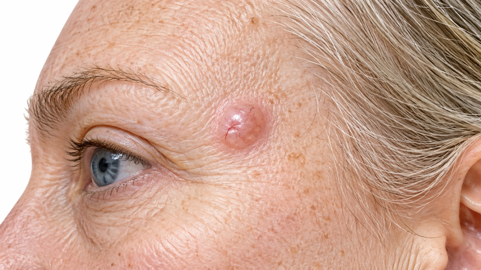
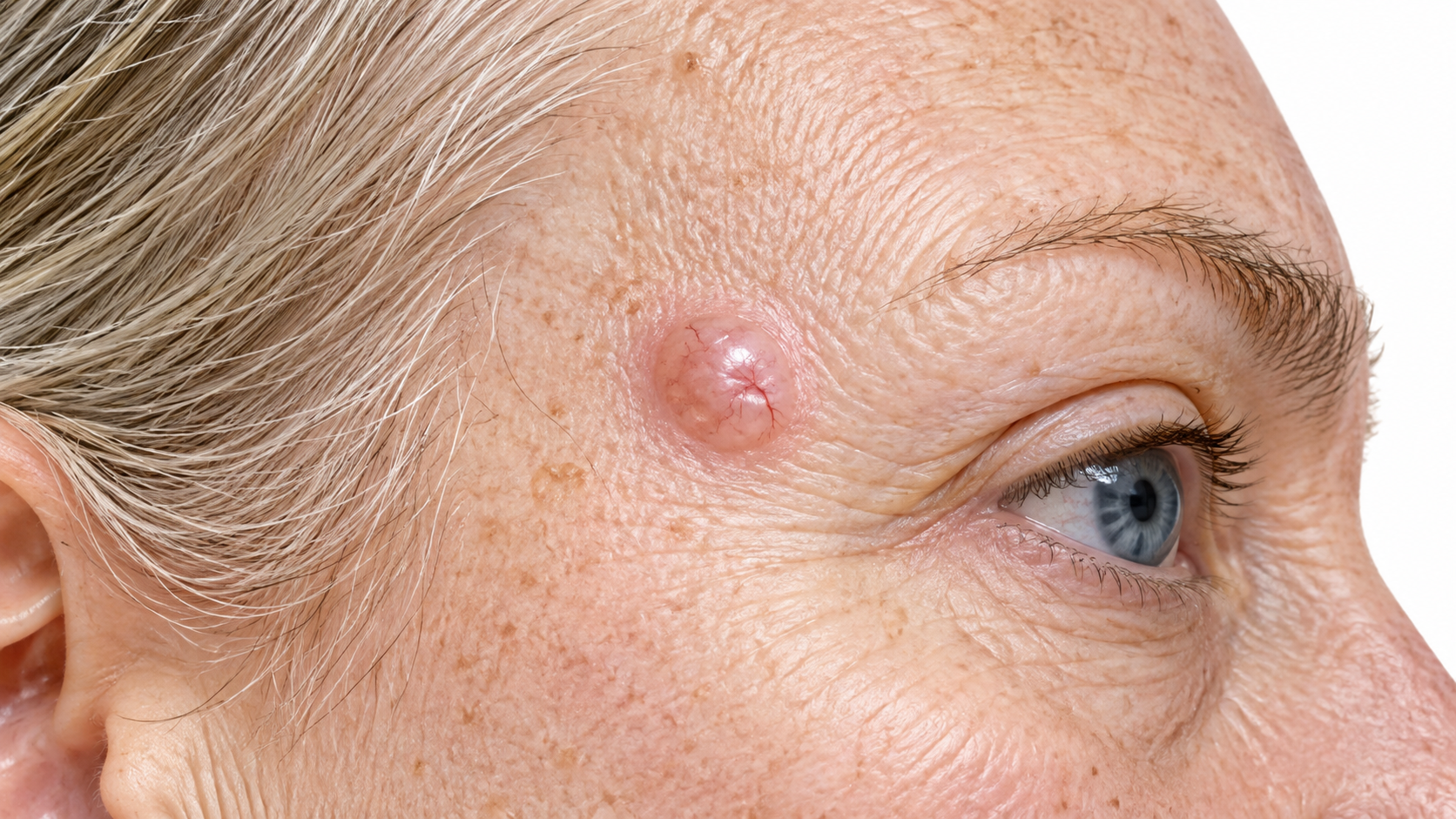
AI-generated educational illustration of Melanoma: 1. Melanoma Header — Light Skin; Lighter skin. Not a clinical photograph.Atypical Moles

Key differences

The same eight facts on every condition comparison.

Usual age
Adults; any age possible
Teens and young adults
Where
Anywhere, including soles and nails
Trunk and limbs
Main sign
A new or changing dark spot
Larger, irregular or multicolored moles
Itch or pain
Usually painless
No
Contagious
No
No
Long term
Curable if removed early
Watched for change
Scars
Removal leaves a scar
No
Who gets it
Fair skin, many moles, sunburns, family history
Fair skin, family history, many moles

What is it?

Melanoma
Atypical Moles
What is it?
  • It is a pigment-cell cancer.
    Melanoma arises from melanocytes, the cells that produce pigment.
  • It can look many ways.
    It may be dark, multicolored, pink or almost unpigmented.
  • Early assessment matters.
    Invasive melanoma can spread beyond the skin.
  • Uncertain spots should not be removed at home.
    Biopsy, not cosmetic removal, is the right step.
  • What it is
    Benign moles with unusual clinical features that can resemble melanoma.

What it looks like

Melanoma

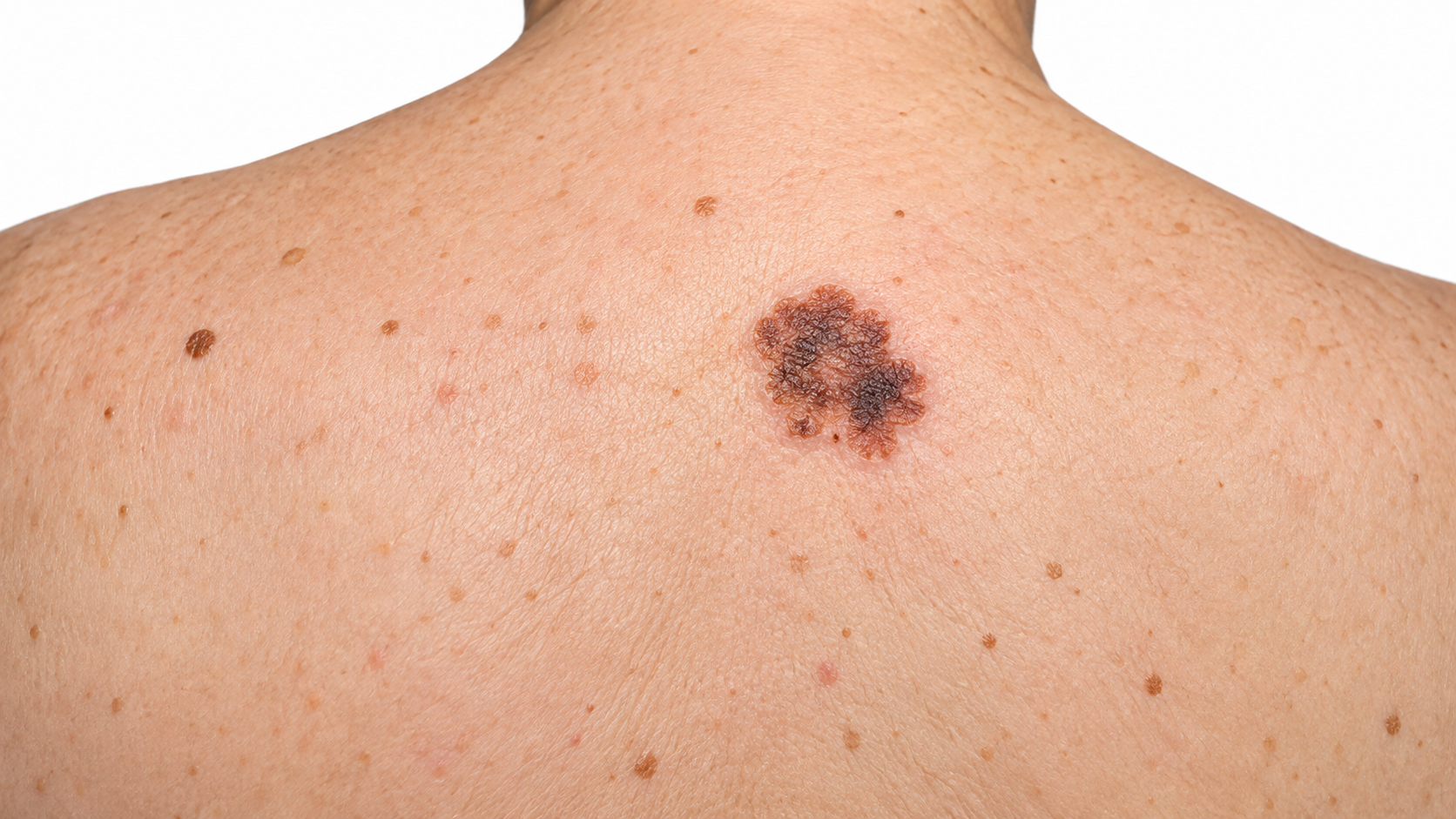
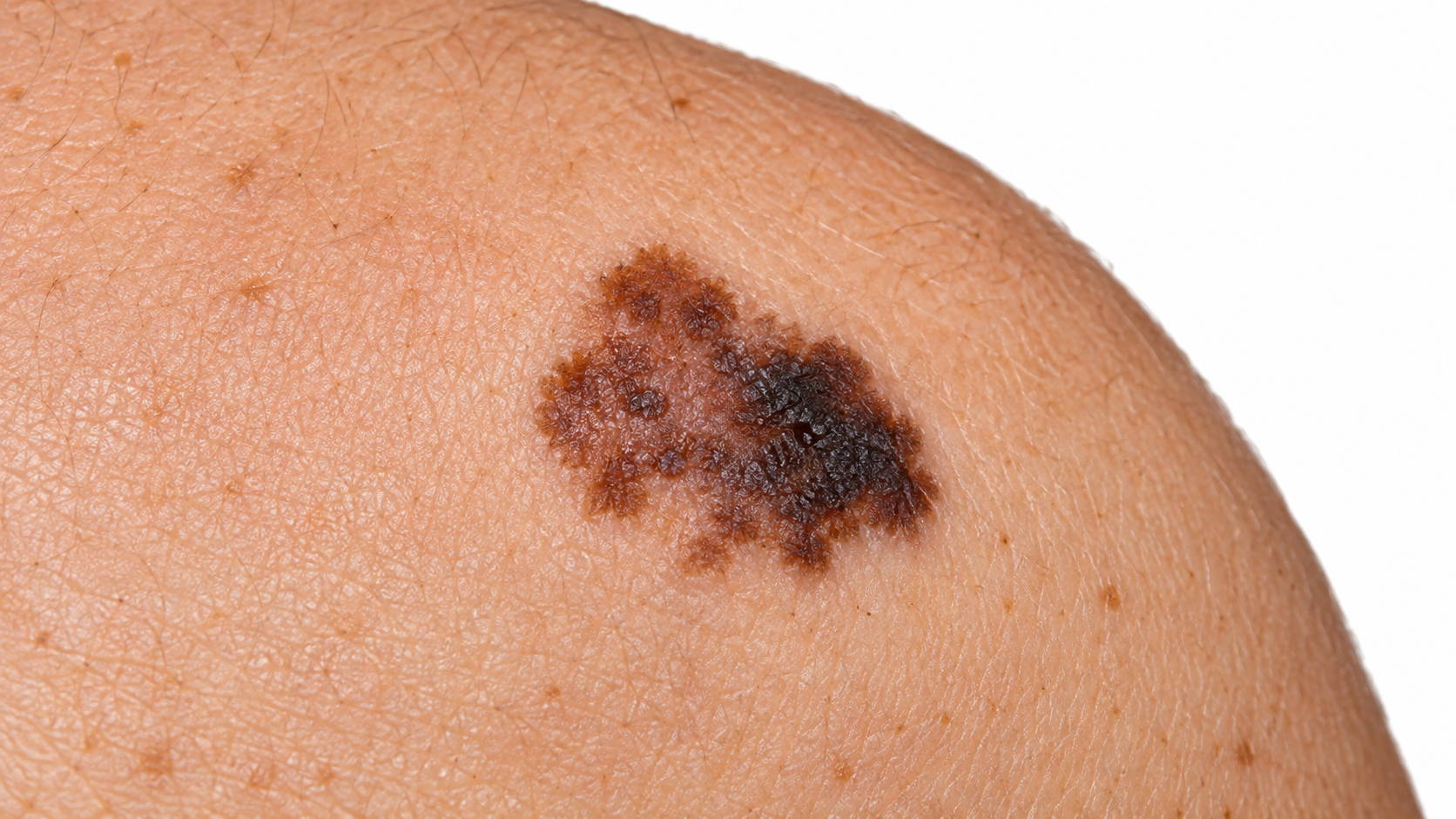
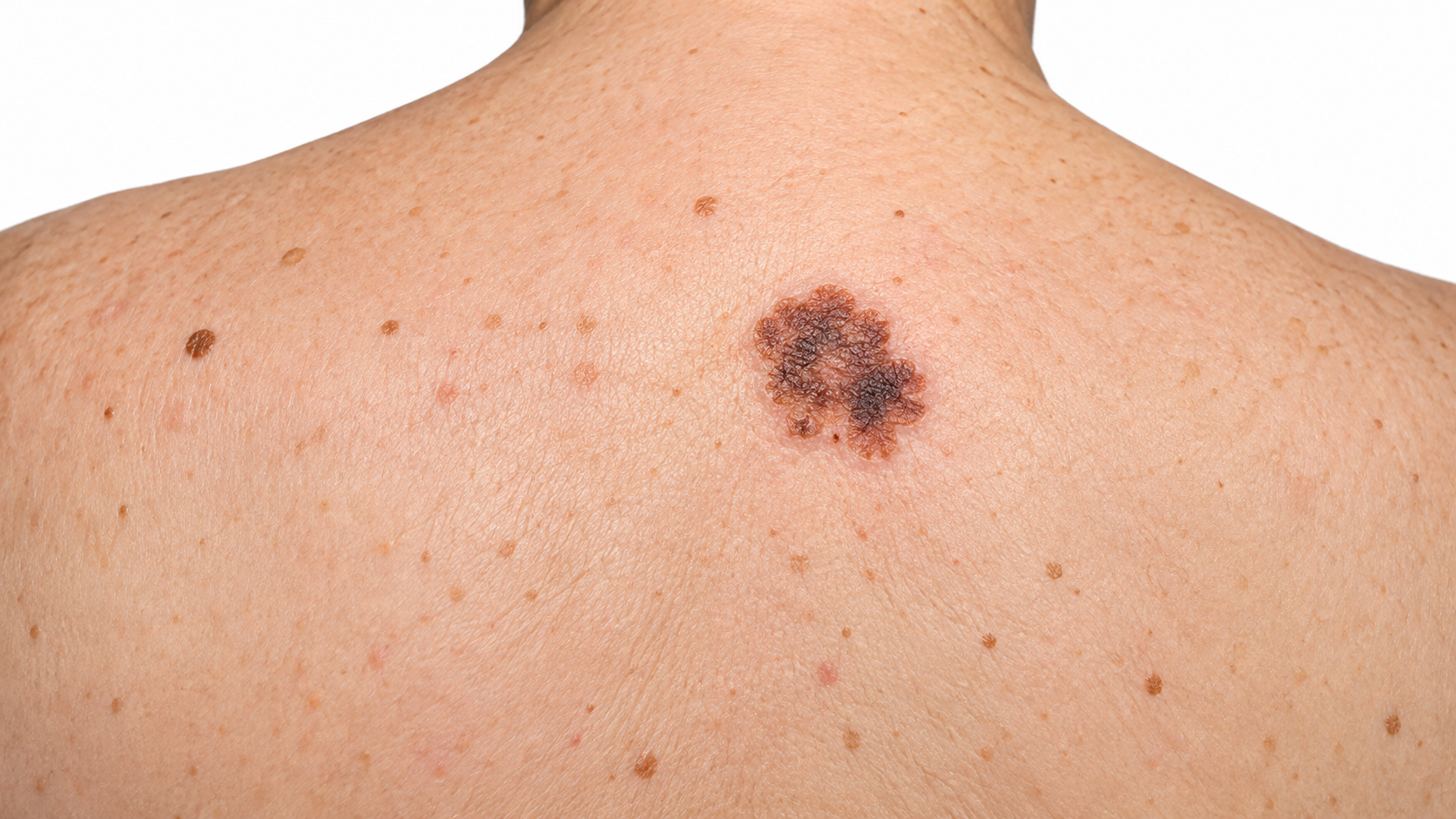
Melanoma: Asymmetry.

Superficial spreading melanoma The most common, a growing flat spot.

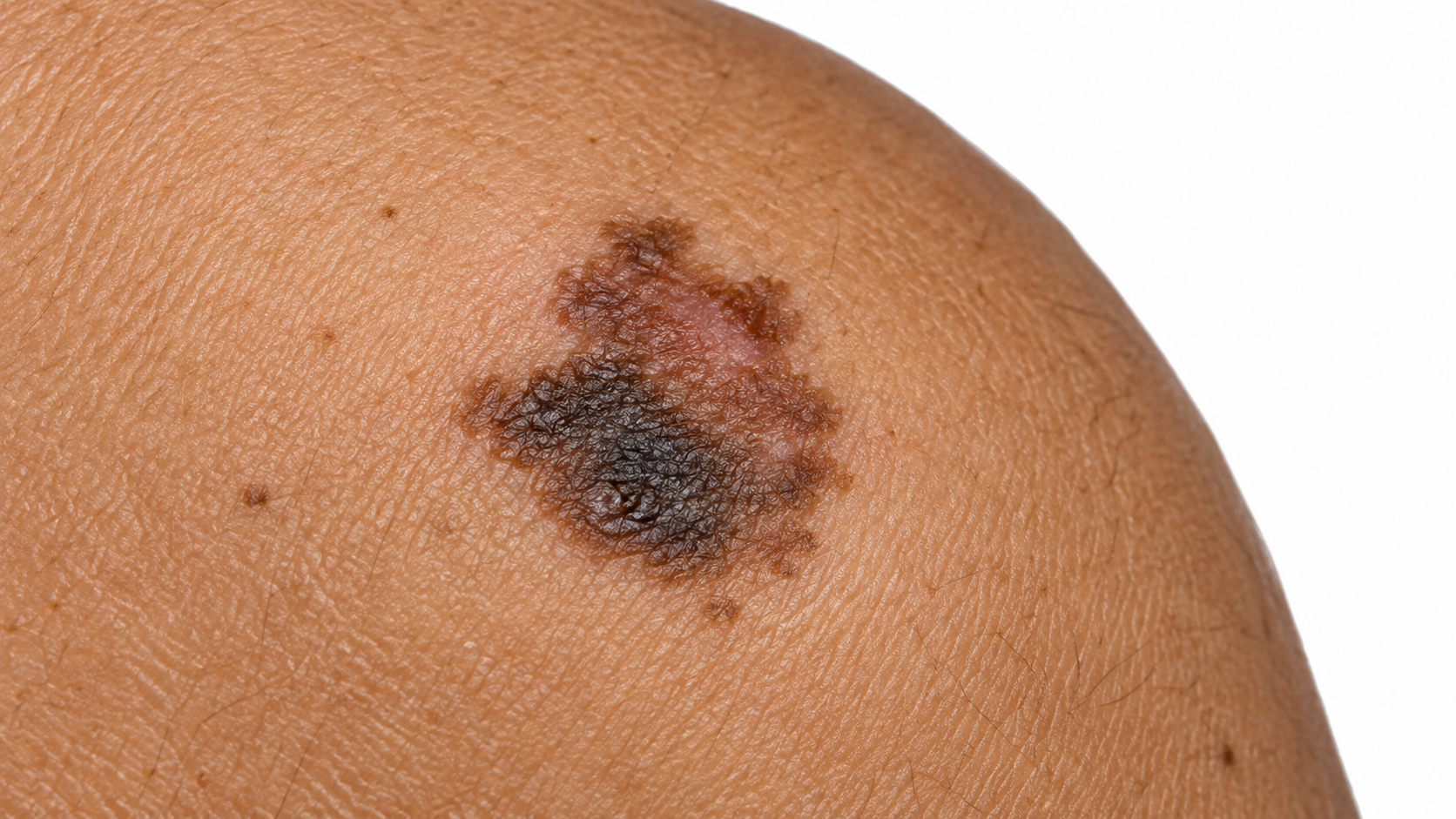
Melanoma: Nodular.

Nodular melanoma A fast-growing raised bump.

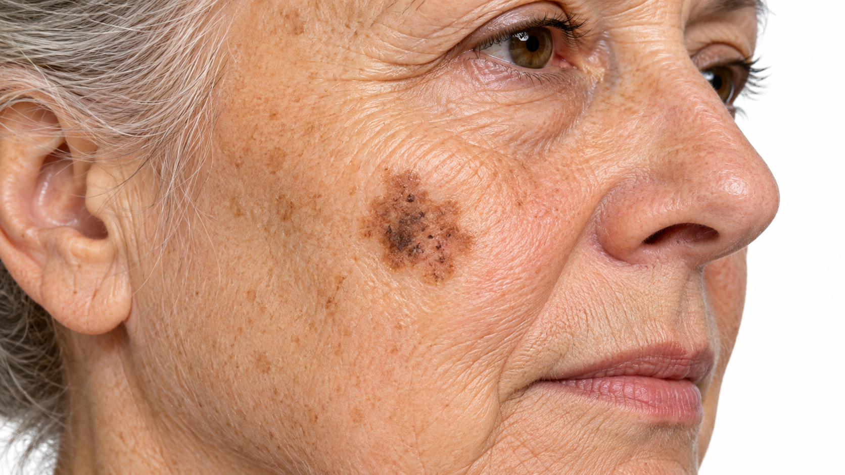
Melanoma: Lentigo maligna.

Lentigo maligna melanoma Slow, flat brown patch on sun-damaged skin.

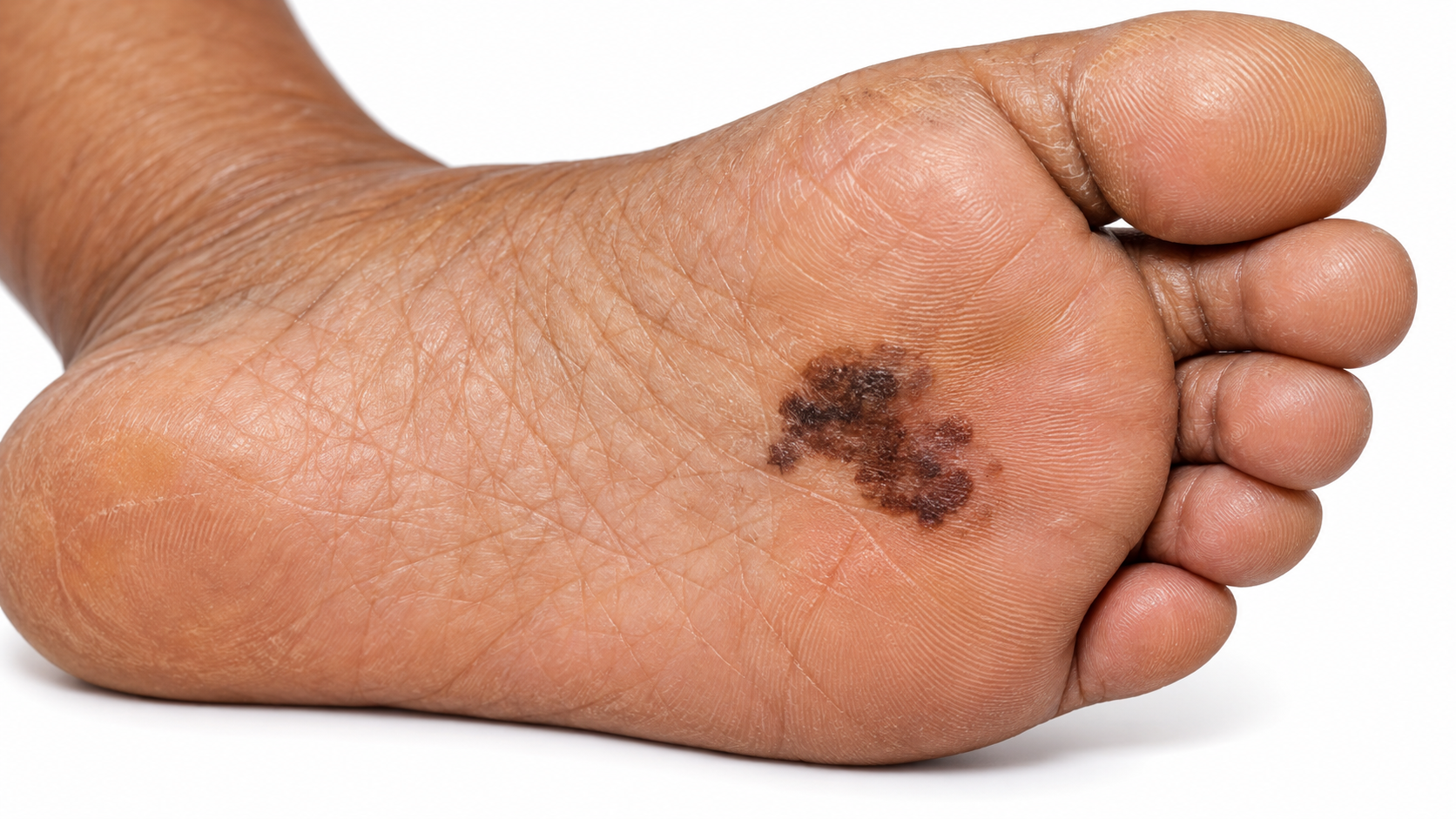
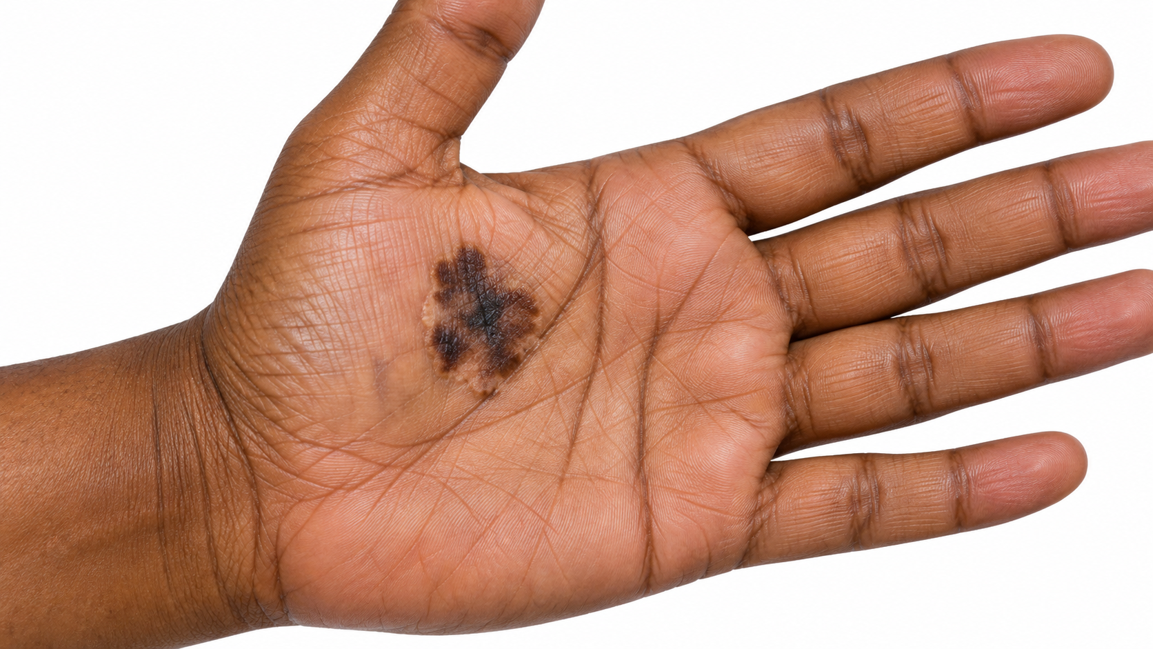
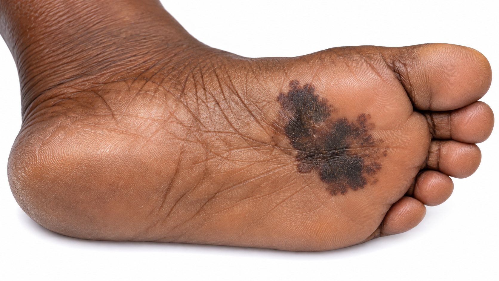
Melanoma: Acral lentiginous.

Acral lentiginous melanoma On palms, soles or under nails.

Atypical Moles

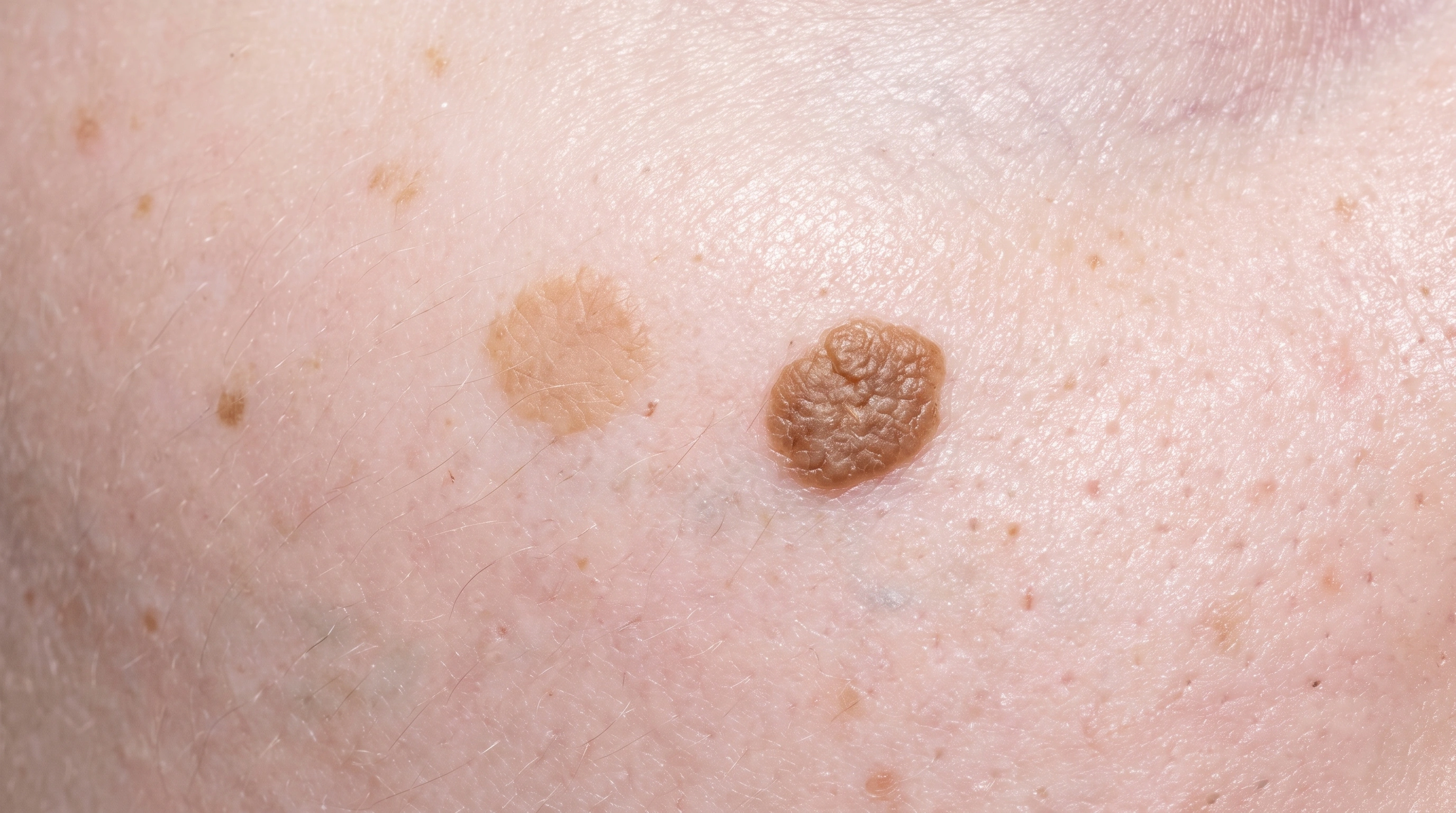
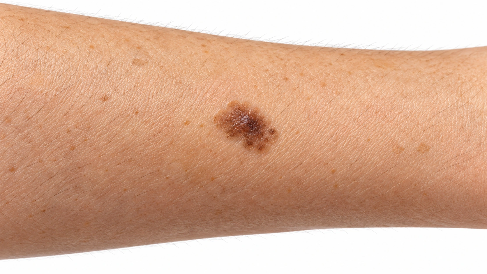
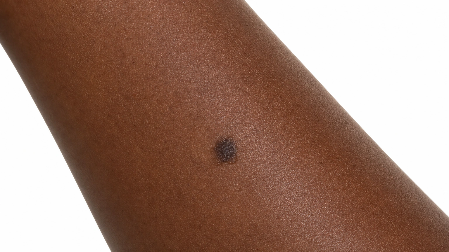
A forearm with an asymmetrical brown spot containing darker areas and uneven borders.

Atypical Moles The pigmented spot has a darker center and an irregular outer edge.

Skin tones
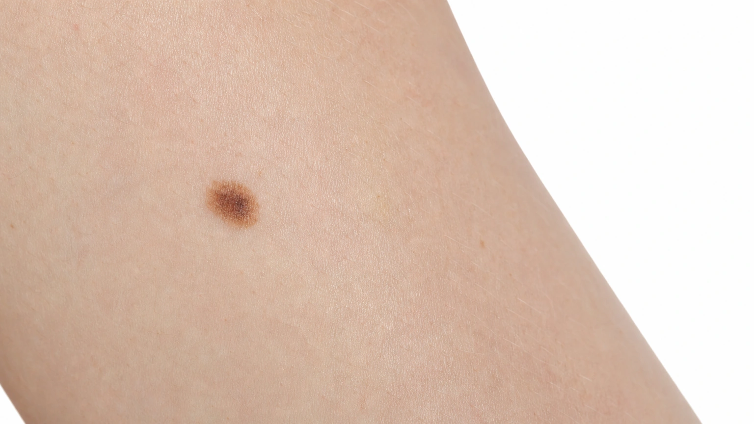
Melanoma: 1. Melanoma Header — Light Skin; Lighter skin.
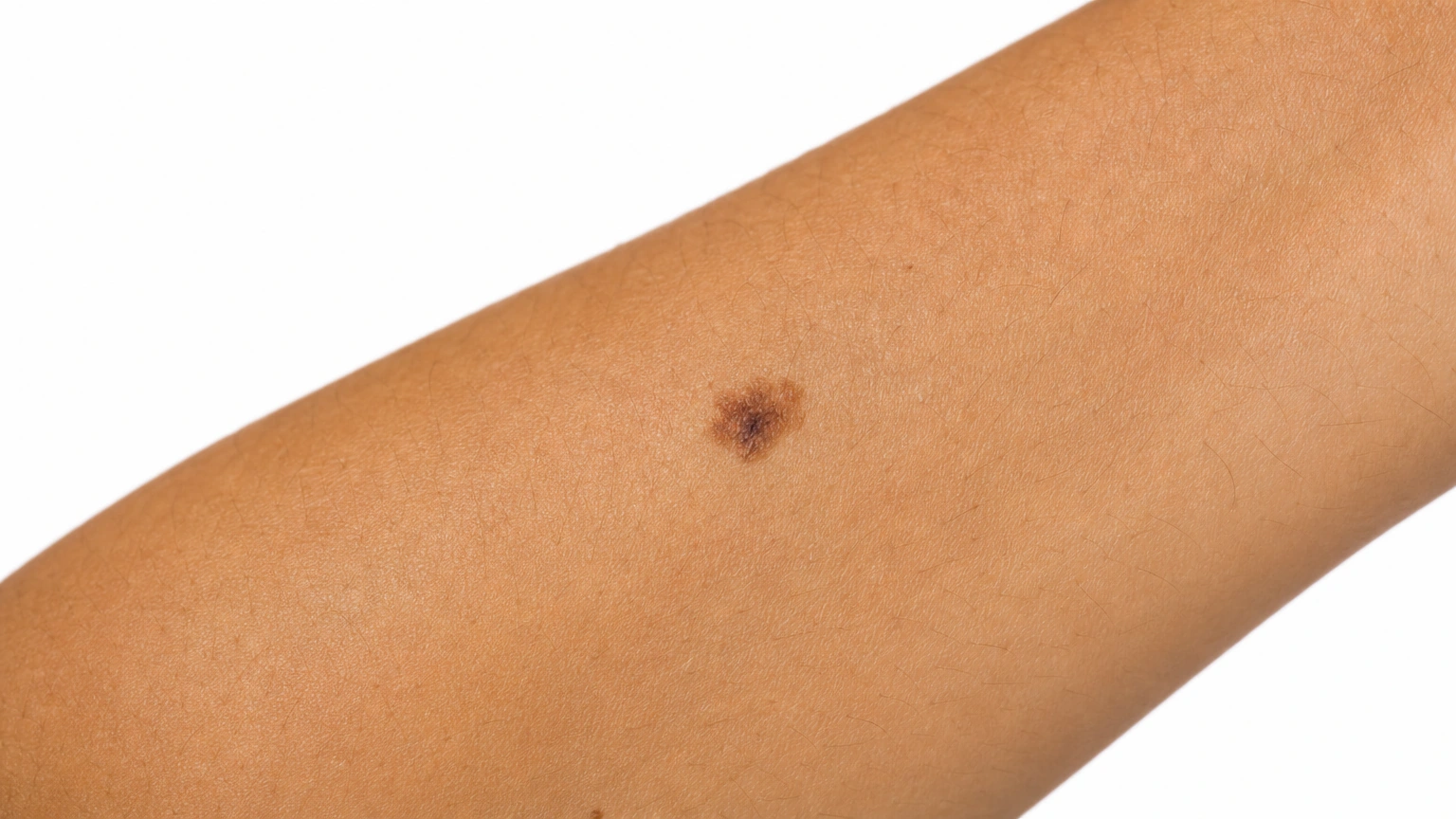
Melanoma: 2. Melanoma Header — Medium Skin; Medium skin.
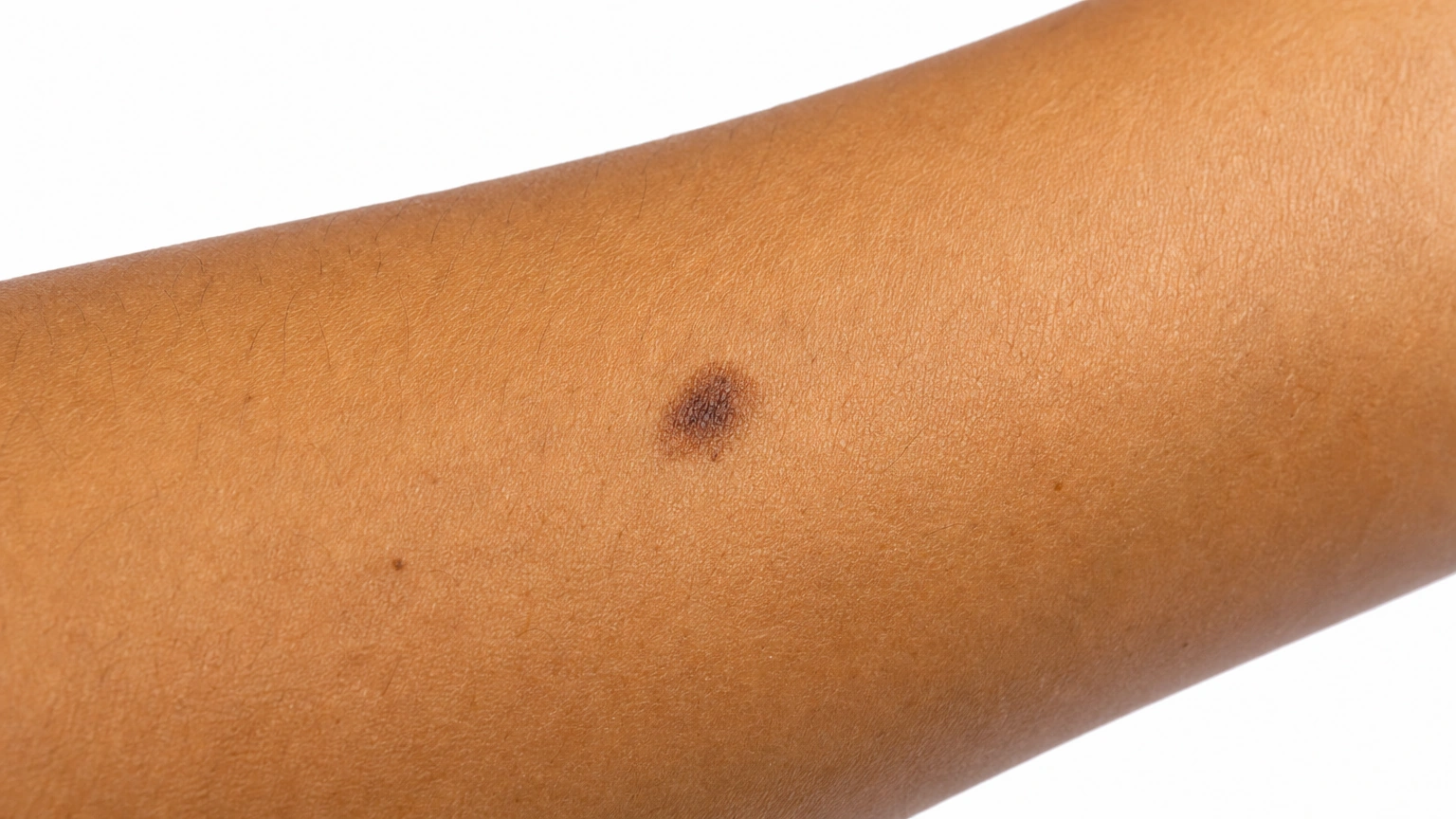
Melanoma: 3. Melanoma Header — Brown Skin; Brown skin.
Melanoma: 4. Melanoma Header — Deep Skin; Darker skin.
a representative asymmetric mole with uneven brown pigment and a mildly irregular border on a forearm on light skin.
a representative asymmetric mole with uneven brown pigment and a mildly irregular border on a forearm on medium skin.
a representative asymmetric mole with uneven brown pigment and a mildly irregular border on a forearm on brown skin.
a representative asymmetric mole with uneven brown pigment and a mildly irregular border on a forearm on deep skin.
Diagram
Melanoma mechanism diagram. Cells multiply unevenly: The patch can become irregular. Pigment cells acquire DNA changes: Abnormal melanocytes escape normal growth control. Some tumors invade more deeply: Depth helps guide staging and treatment.
In the skin

Abnormal pigment cells escape normal growth controls and multiply within the skin. Some melanomas grow into deeper tissue, and the depth of invasion helps determine their stage.

Benign pigment-cell growth can produce a mole with an unusual visible appearance. Surface appearance and microscopic findings are different assessments, so atypical appearance alone does not establish cancer.

When it usually starts

010203040506070+

Melanoma. More common in adults, but a concerning changing lesion needs assessment at any age.

Atypical Moles. Often develops during adolescence or early adult life.

Causes and triggers

Causes

Melanoma starts when pigment cells develop DNA damage and grow out of control. Ultraviolet light is the main cause of that damage for most melanomas.

A benign melanocyte proliferation has an atypical appearance; clinical appearance and pathology are different assessments.

Family tendency

About 1 in 10 people with melanoma have a family history of it. Genes such as CDKN2A raise the risk.

Risk factors
Fair skin and red or blond hair, Many moles, Past blistering sunburns, Weakened immunity, Family history
Family tendency, Many moles, Personal melanoma history
Trigger
● Melanoma
● Atypical Moles
Sunburns
Well established
Not listed
Tanning beds
Well established
Not listed
Intense, on-and-off sun
Strong link
Not listed
No established short-term flare trigger
Not listed
Not a typical trigger

Daily Care

Melanoma
Atypical Moles
Daily care

Habits that help

  • Learning the ABCDE signs
    Asymmetry, Border, Color, Diameter, Evolving above all.
  • The ugly duckling rule
    The mole that does not belong is worth examining.
  • Knowing ABCDE is not complete
    Firm, growing and new counts on its own.
  • Checking the places nobody checks
    Scalp, ears, soles, between the toes, nails, genitals and mouth.
  • Photographing your own skin
    Same distance and light, every few months.
  • Daily sun protection, no tanning beds
    Cuts future risk, and you control it.
  • Review immune-suppressing medicines with the prescriber; do not stop them yourself
    Immune suppression can change skin-cancer risk and follow-up needs. Do not stop a prescribed medicine without the specialist’s plan.

Avoid

  • Avoid: Tanning beds
    A recognized cause, riskier the younger you start.
  • Avoid: Blistering sunburns in childhood
    Linked more strongly than steady daily exposure.
  • Avoid: Only checking sun-exposed areas
    The scalp, soles, nails and mouth are found late.
  • Avoid: Assuming a pink bump is harmless
    Melanoma without pigment is a recognized variant.
  • Avoid: A fast-growing bump ignored
    Nodular melanoma grows downward from the start.
  • Avoid: Wearing sunscreen only on holiday
    Risk builds over a lifetime of ordinary days.
  • Avoid: Waiting out a changing spot
    A melanoma watched for six months is thicker.
  • Avoid: Treating a spot at home
    Freezing kits and black salve destroy the evidence.
  • Avoid: Skipping follow-up
    A second melanoma is meaningfully more likely.
  • Avoid: Not telling close relatives
    It changes their screening advice.
  • Avoid: Cryotherapy or cosmetic laser
    Freezing or lasering a pigmented spot off destroys it without anyone ever finding out what it was. If it was a melanoma, the thickness can no longer be measured and no one can check the edges. A spot that concerns you is examined and sampled, not erased for convenience, and this is one of the few places where the cheaper, quicker option costs something real.

Habits that help

  • Use broad-spectrum SPF 30 or higher on exposed skin
    Reapply during outdoor exposure and add shade, a hat and protective clothing.
  • Know your usual skin pattern and notice new changes
    Look for a spot that changes or stands out from the rest rather than checking size alone.
  • Take clear comparison photographs when advised
    Consistent lighting can help document a change over time.
  • Keep any recommended skin-examination appointments
    Frequency depends on personal and family history and the clinician’s advice.

Avoid

  • Avoid: Tanning beds and deliberate sun tanning
    UV damage increases skin cancer risk.
  • Avoid: Home acids, freezing or cutting pigmented spots
    A changing lesion needs assessment before cosmetic removal.

Treatment differences

Evidence labels come from each condition’s own treatment list.

Treatment
● Melanoma
● Atypical Moles
Procedures
Editorial illustration for Surgical Excision, based on the entry’s treatment or equipment reference.

Surgical Excision

Strong evidence

The treatment that cures the great majority of thin melanomas. Once the diagnosis is confirmed, a second and wider operation removes a margin of surrounding skin, with the width set by how thick the melanoma was. The scar is longer than most people expect, and that is the trade that stops it coming back at the site. Anything pigmented that is removed, anywhere, should be sent for pathology.
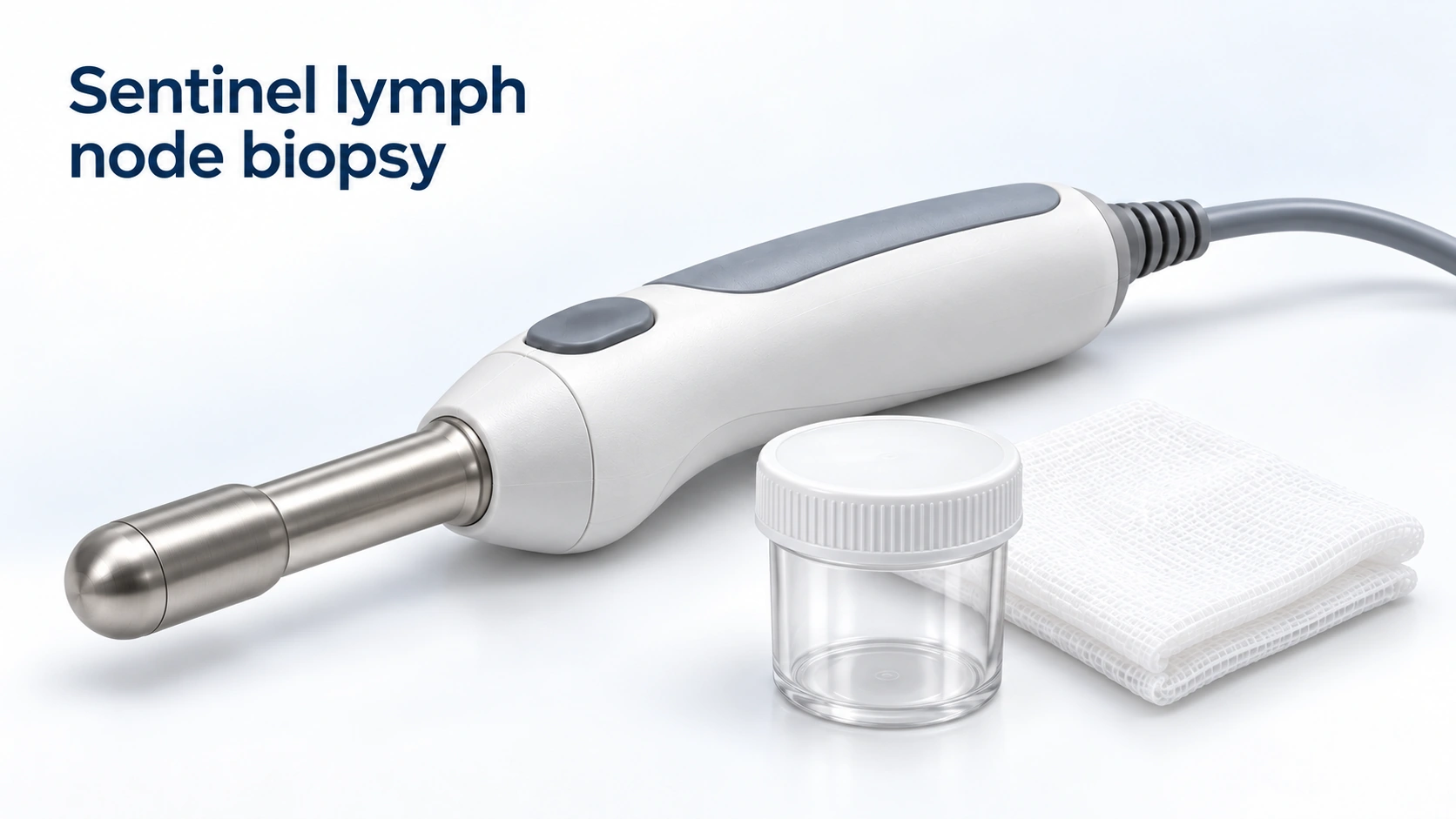
Editorial illustration for Sentinel lymph node biopsy, based on the entry’s treatment or equipment reference.

Sentinel lymph node biopsy

Strong evidence

Above a certain thickness, the first lymph node the area drains to is located with a tracer and removed to see whether cells have reached it. It is a staging test rather than a treatment: it tells you where you stand and shapes what is offered next. It carries its own risks, including lasting swelling of the limb, so it is offered as a discussion rather than an automatic step.

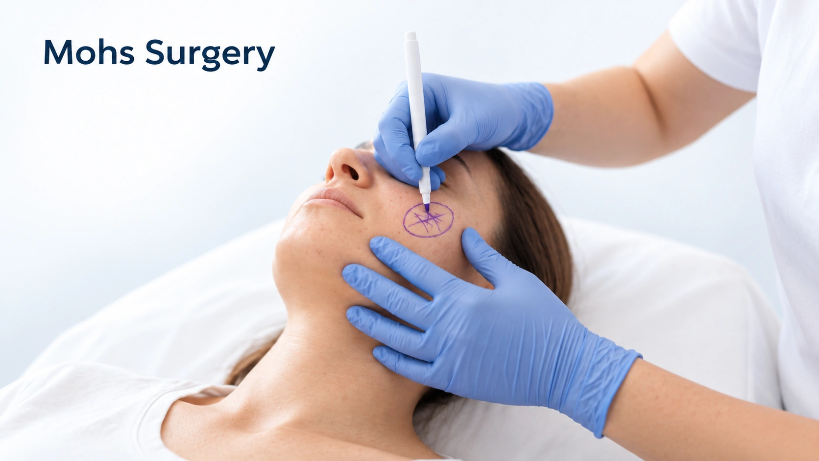
Editorial illustration for Mohs Surgery, based on the entry’s treatment or equipment reference.

Mohs Surgery

Moderate evidence

Used at some centers for lentigo maligna on the face, where sparing skin matters and the true edge of the pigment is hard to see. It is not the standard operation for melanoma generally — wide excision is, and the evidence sits with that. If Mohs is offered to you, it is fair to ask why it suits this particular lesion in this particular place.

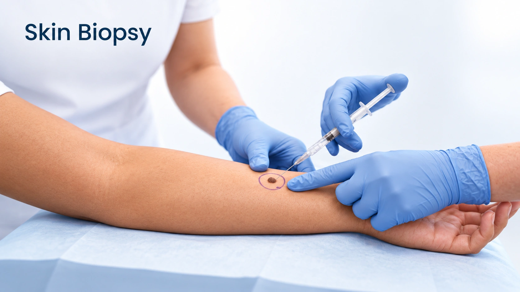
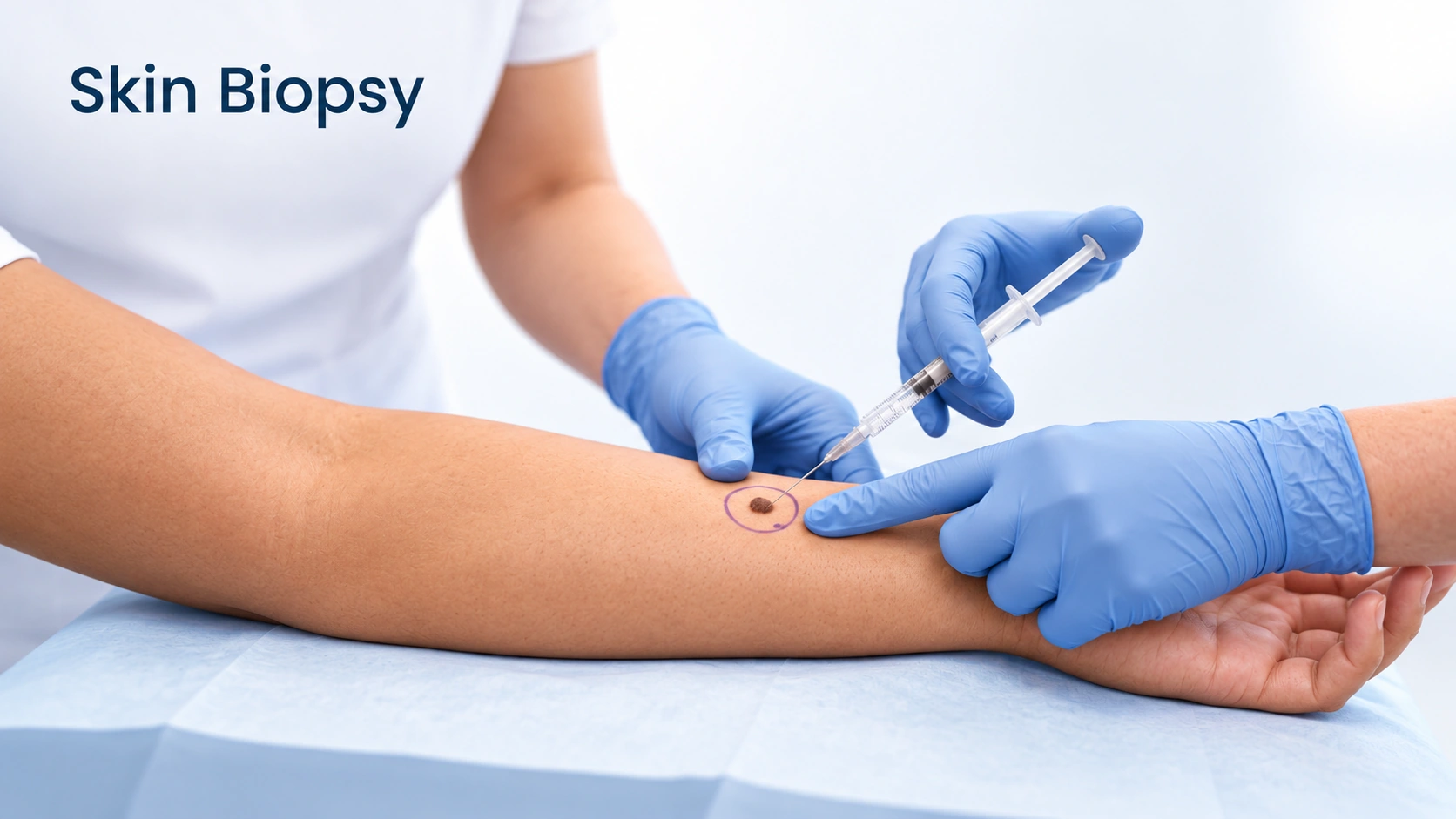
Editorial illustration for Skin Biopsy, based on the entry’s treatment or equipment reference.

Skin Biopsy

Listed

Nothing diagnoses melanoma except a biopsy. Where it is possible, the whole spot is removed with a narrow margin rather than sampled, because the pathologist needs the full depth to measure the thickness, and thickness sets everything that follows. It is done under local anesthetic and usually takes under half an hour. A biopsy does not make melanoma spread, and believing that it does is a common cause of delay.

Biopsy of concerning lesions

Listed

Atypical appearance alone does not require removing every mole; suspicious change may need tissue diagnosis.

Outlook

Long term

Melanoma does not progress on a fixed schedule. Some variants change over years, others over weeks, so this is the sequence, not a timetable. Skin checks continue for years after treatment, because a second melanoma and a recurrence are both possible, and close relatives are usually advised to have their skin checked.

Most atypical moles remain benign, but their presence can indicate a higher melanoma risk. Monitoring follows personal risk; a changing mole needs examination rather than reassurance based on its label.

Complications

Possible complications
  • Cells travel to the nearest nodes first
  • Treated by an oncology team
More details

Cells travel to the nearest nodes first
A lump near a previous melanoma site is significant and checked promptly.

Treated by an oncology team
Melanoma can spread to organs far from the skin. Immunotherapy and targeted therapy have changed outcomes substantially in the last decade.

Multiple atypical moles can mark increased melanoma risk; not every one requires removal.

When to get help

When to get help

Arrange prompt assessment of a new or changing unusual spot, especially an evolving lesion or one that bleeds. Diagnosis requires clinical evaluation and usually biopsy.

Seek prompt assessment for a changing mole, an unusual new lesion or a spot unlike your others.

Common questions

Melanoma:How quickly does melanoma grow?

It varies more than people expect. The common superficial spreading variant often changes over months to a few years. Nodular melanoma can grow downward over weeks to a few months. You cannot tell which you have by looking, so a changing spot gets examined now, not in six months.

Melanoma:Does having a mole removed spread cancer?

No, and this belief causes delay. Removing a suspicious lesion is how melanoma is diagnosed and how the thickness is measured, and thickness determines the treatment. There is no evidence a biopsy spreads it.

Melanoma:Should I have all my moles removed to be safe?

No. Most melanomas arise as new spots, so removing moles does not remove the risk and means many scars for no benefit. Many or unusual moles are a reason for regular examinations, not preventive removal.

Melanoma:I have a dark line on my nail. Is that melanoma?

Usually not. Pigmented nail bands are common and often normal in deeper skin tones, frequently on several nails at once. It needs examining if it is on one nail only, if it is getting wider, if it has more than one shade, if the nail is splitting, or if the pigment spreads onto the skin around it.

Melanoma:If it is removed and the margins are clear, am I finished?

The melanoma is treated, but you are not discharged. Having had one raises the chance of a second, so skin checks continue for years, and close relatives are usually advised to have their skin examined. Ask how often yours should be and who arranges it.

Melanoma:I never sunbathe. Do I still need to worry about a changing spot?

Yes. Sun exposure raises risk, but melanoma also occurs on soles, palms, under nails and on skin never exposed, and in people who have never burned. Sun history changes your risk profile, not whether a particular spot should be looked at.

Atypical Moles:What is Atypical Moles?

Benign moles with unusual clinical features that can resemble melanoma.

Atypical Moles:What can I do day to day?

Avoid picking or home removal. Protect exposed skin from the sun.

Atypical Moles:When should I get medical advice?

Seek prompt assessment for a changing mole, an unusual new lesion or a spot unlike your others.

Sources

Sources and further reading

  1. DermNet
  2. Atypical melanocytic naevus
Dermapedia on YouTube