Melanoma vs Seborrheic Keratosis

Melanoma is a skin cancer that starts in the cells that make pigment. It is much less common than other skin cancers but causes most skin cancer deaths, because it can spread. Found and removed while it is still thin, it is usually curable. A seborrheic keratosis is a common, harmless growth that looks stuck onto the skin, like a piece of wax or a small barnacle. They are not cancer and they do not become cancer. The reason to see a doctor is to be sure that is what you are looking at.

Reviewed by Dr. Schwarz, Board-Certified Dermatologist · Research updated 2026-10-23
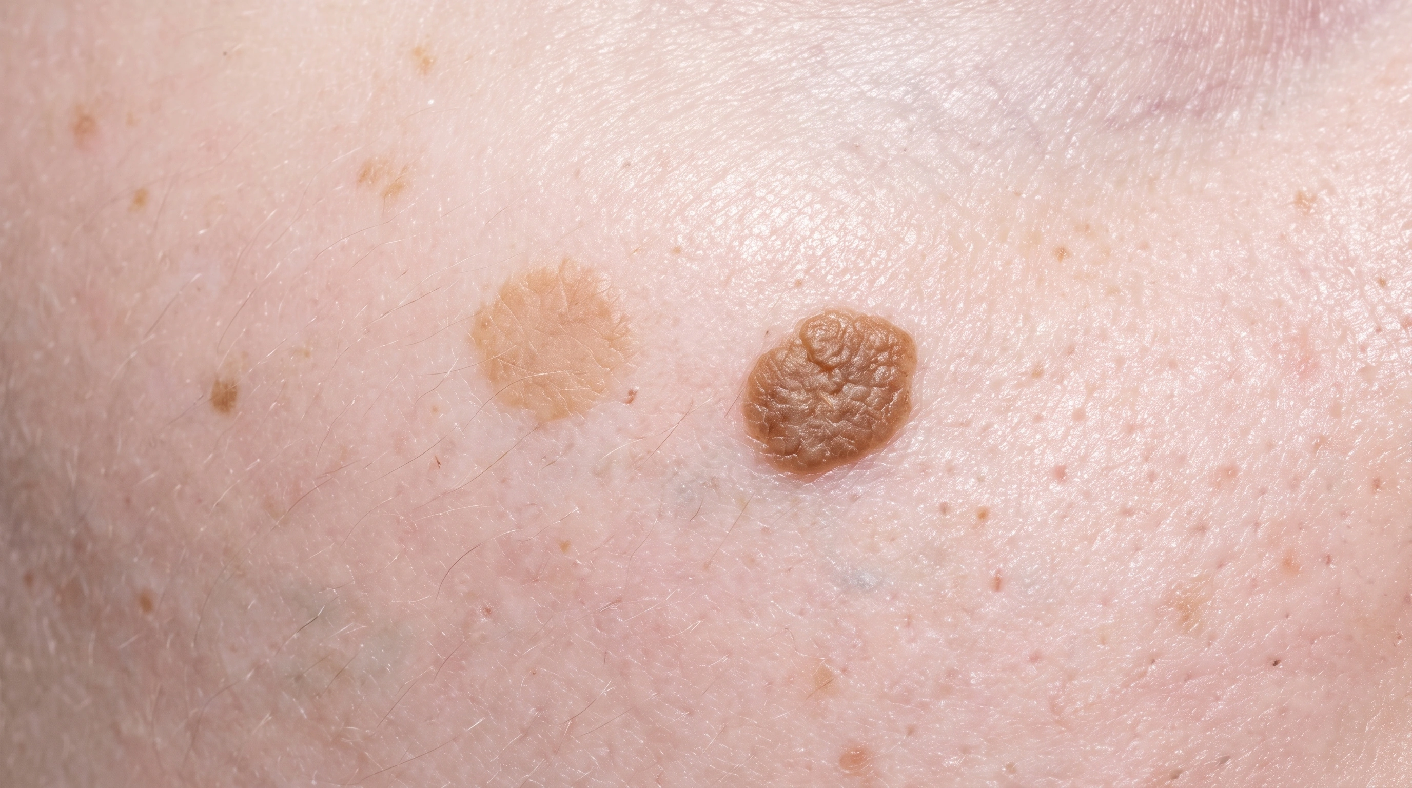
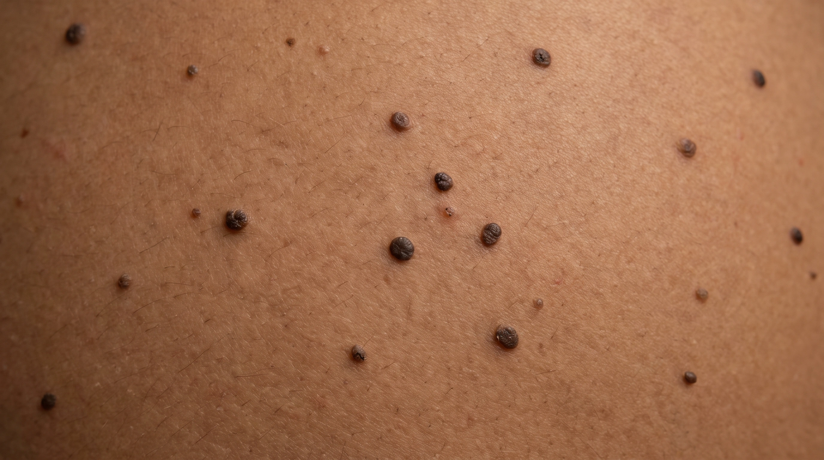
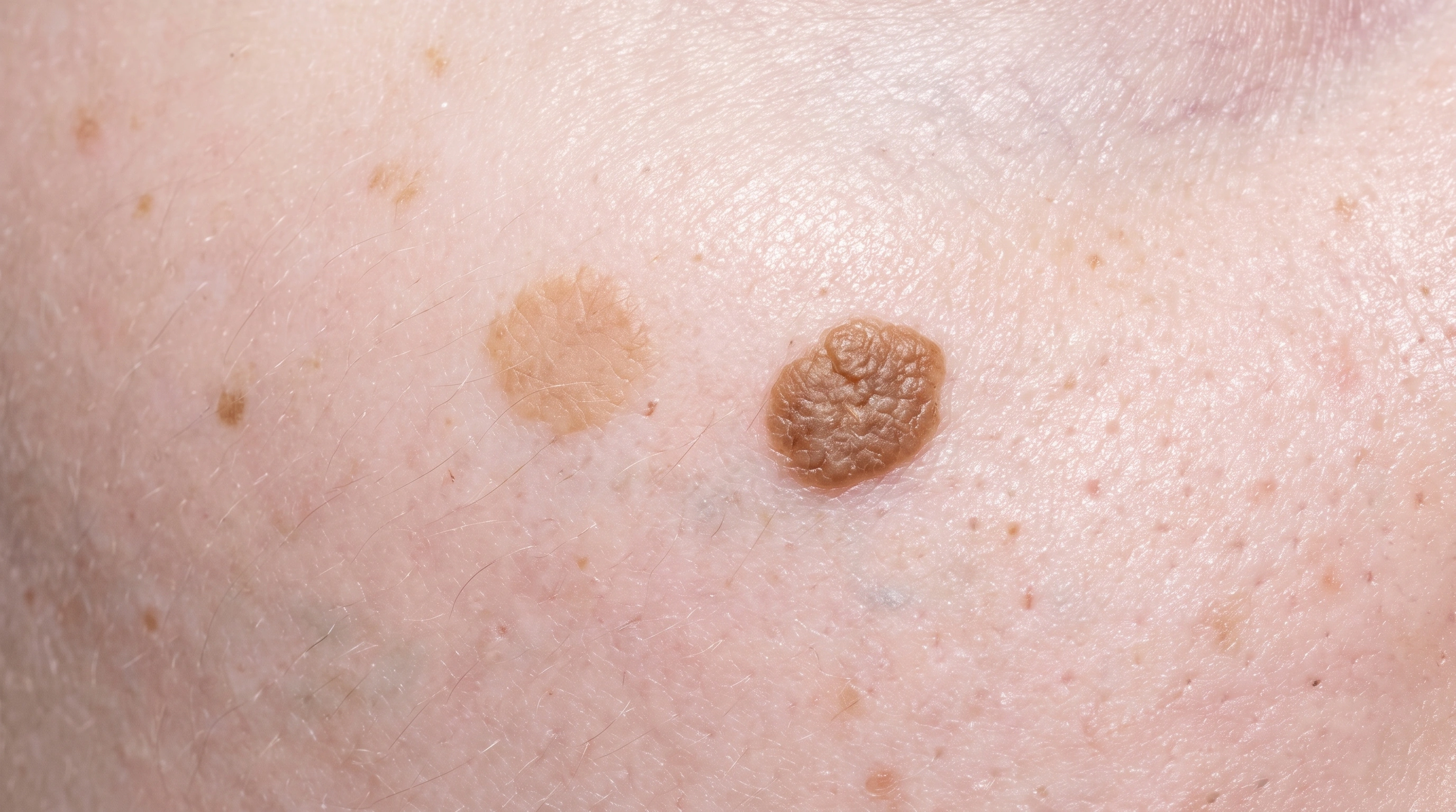
AI-generated educational illustration of Melanoma: 1. Melanoma Header — Light Skin; Lighter skin. Not a clinical photograph.Two growths on the upper back on fair skin. One is a flat light tan patch and the other is a raised mid-brown growth with a rough waxy surface and a sharply defined edge.

Key differences

The same eight facts on every condition comparison.

Usual age
Adults; any age possible
40 and over
Where
Anywhere, including soles and nails
Back, chest, face
Main sign
A new or changing dark spot
Waxy stuck-on growth
Itch or pain
Usually painless
Sometimes itchy
Contagious
No
No
Long term
Curable if removed early
Stays; more appear
Scars
Removal leaves a scar
No
Who gets it
Fair skin, many moles, sunburns, family history
Most people over 50

What is it?

Melanoma
Seborrheic Keratosis
What is it?

It is a pigment-cell cancer.
Melanoma arises from melanocytes, the cells that produce pigment.

It can look many ways.
It may be dark, multicolored, pink or almost unpigmented.

Early assessment matters.
Invasive melanoma can spread beyond the skin.

Uncertain spots should not be removed at home.
Biopsy, not cosmetic removal, is the right step.

It is a benign growth.
Surface skin cells form a waxy, warty or stuck-on growth.

Color varies.
It can be tan, brown, gray or almost black.

It is not a precancer.
These growths become more common with age.

Uncertain lesions are tested.
Tissue is examined before any destructive treatment if the appearance is unclear.

What it looks like

Melanoma

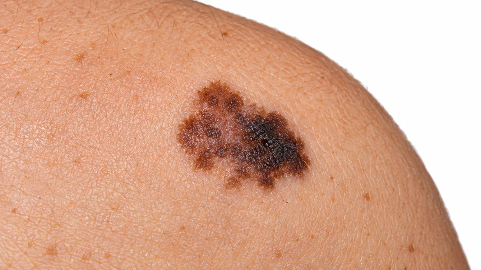
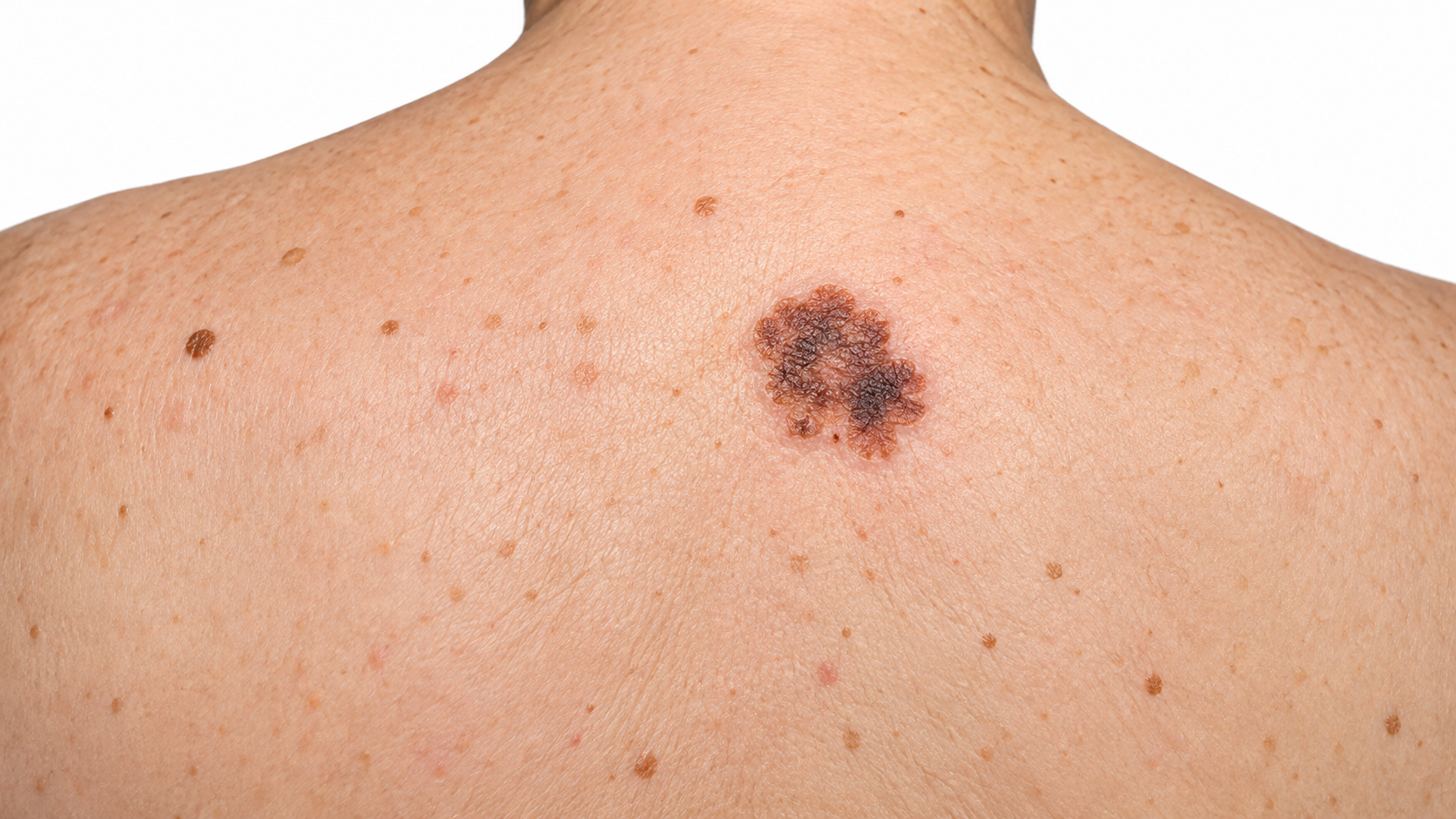
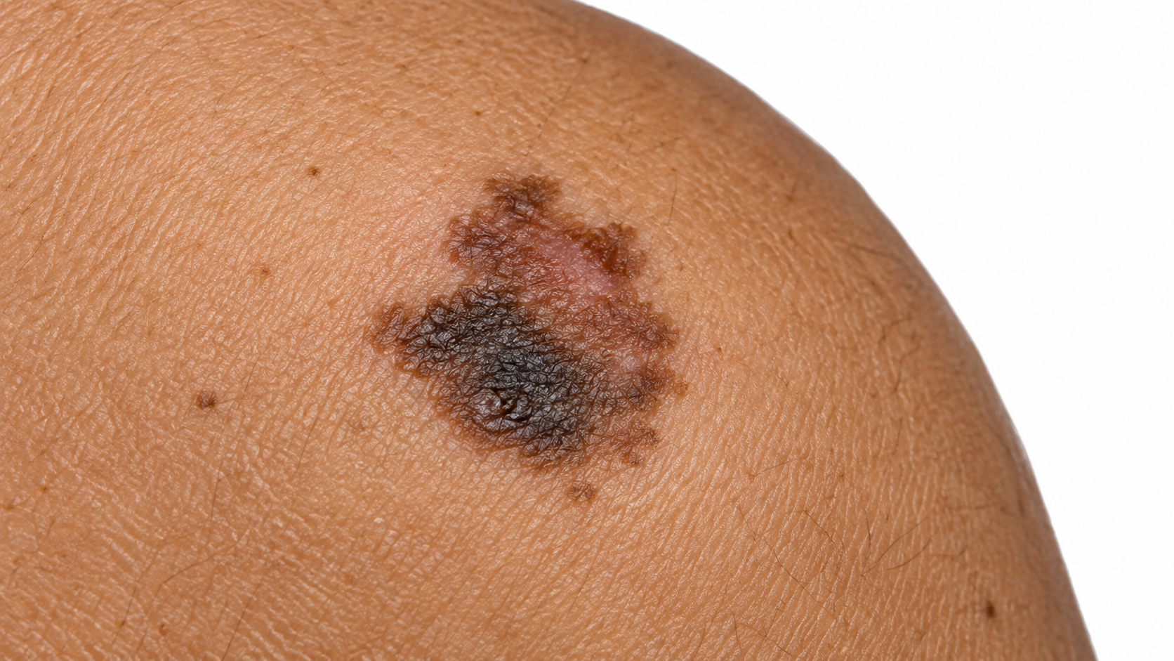
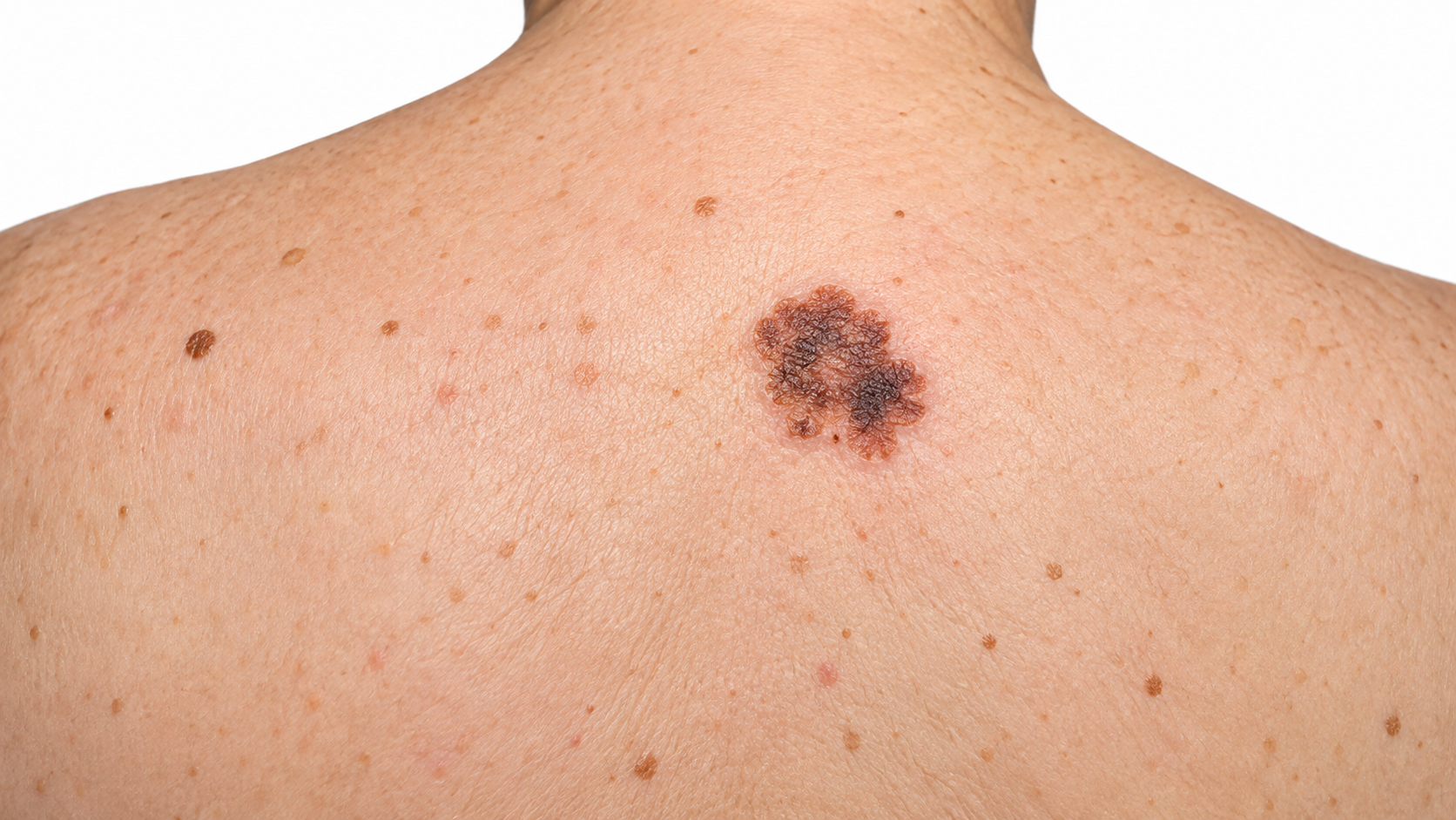
Melanoma: Asymmetry.

Superficial spreading melanoma The most common, a growing flat spot.

Melanoma: Nodular.

Nodular melanoma A fast-growing raised bump.

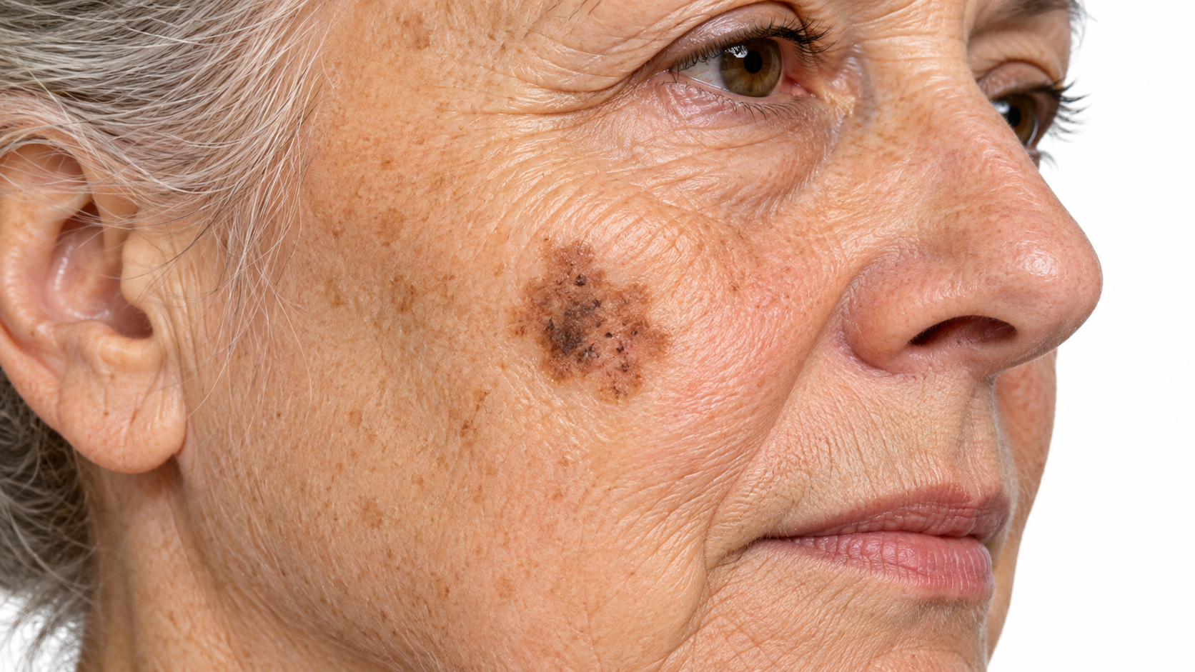
Melanoma: Lentigo maligna.

Lentigo maligna melanoma Slow, flat brown patch on sun-damaged skin.

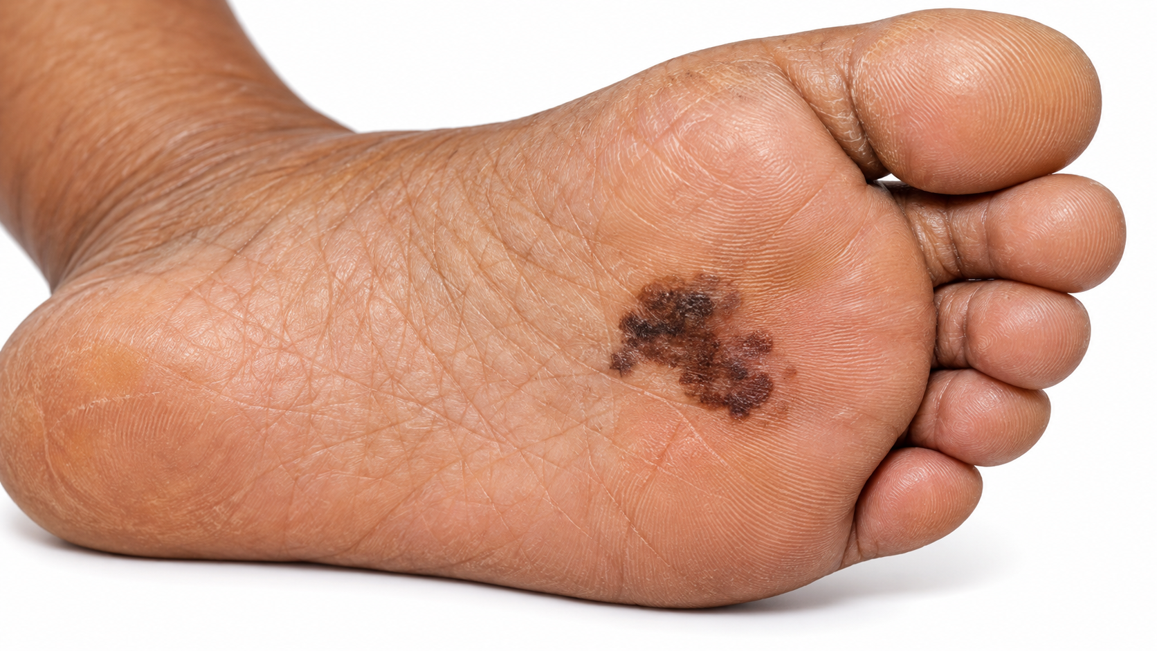
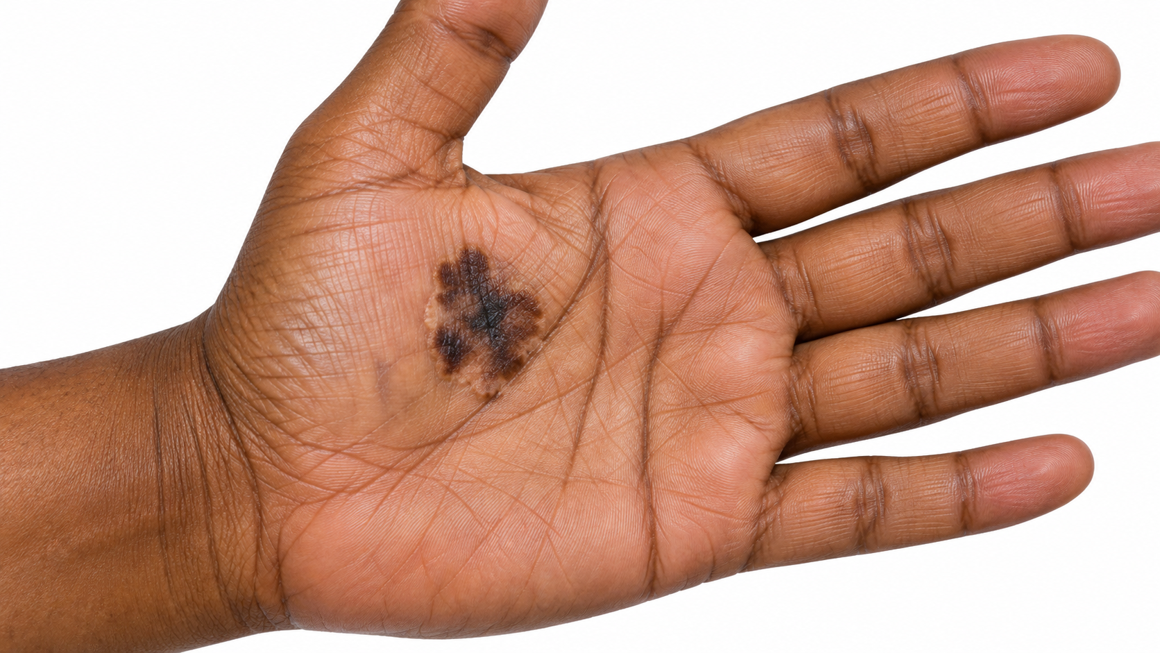
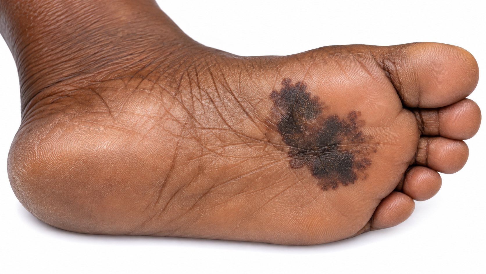
Melanoma: Acral lentiginous.

Acral lentiginous melanoma On palms, soles or under nails.

Seborrheic Keratosis

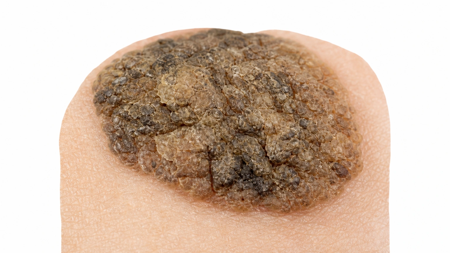
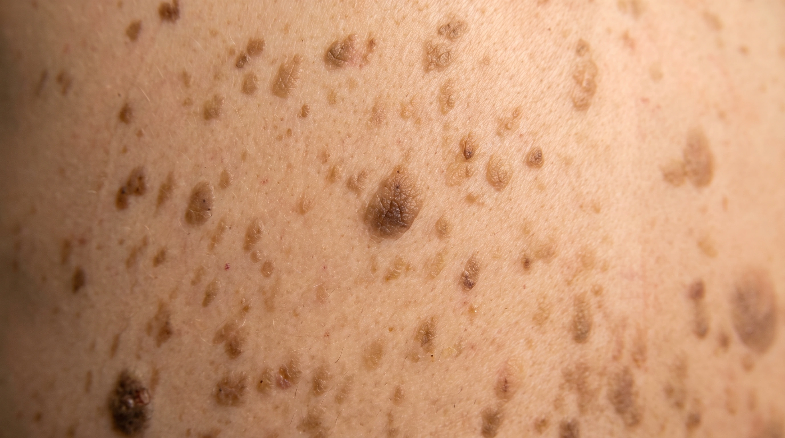
Close view of a rough hyperkeratotic brown stuck-on lesion, representing seborrheic keratosis.

Classic seborrheic keratosis Raised, waxy, stuck-on growth.

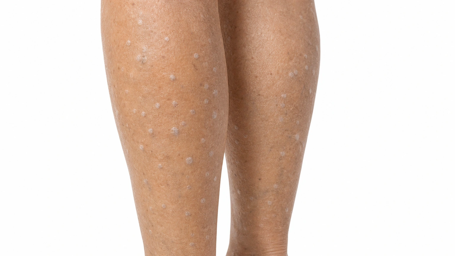
Multiple small gray-white raised bumps on the lower legs, representing stucco keratoses.

Stucco keratosis Small white-gray bumps on the legs.

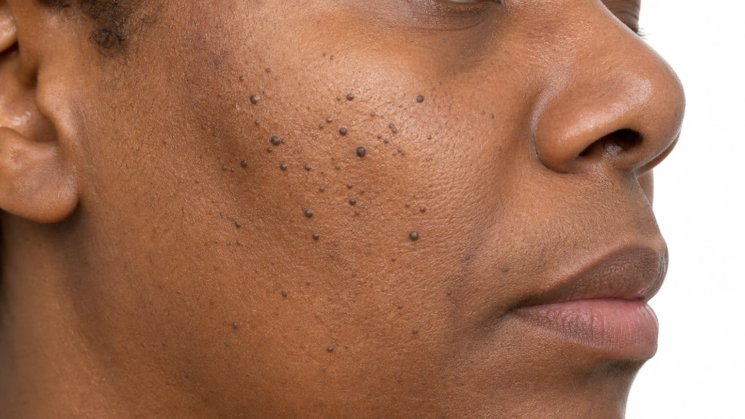
Small dark raised papules on brown cheek skin, representing dermatosis papulosa nigra.

Dermatosis papulosa nigra Small dark bumps on the face.

Skin tones
Melanoma: 1. Melanoma Header — Light Skin; Lighter skin.
Melanoma: 2. Melanoma Header — Medium Skin; Medium skin.
Melanoma: 3. Melanoma Header — Brown Skin; Brown skin.
Melanoma: 4. Melanoma Header — Deep Skin; Darker skin.
Two growths on the upper back on fair skin. One is a flat light tan patch and the other is a raised mid-brown growth with a rough waxy surface and a sharply defined edge.
A raised brown growth on the chest on medium olive skin. Its color is only slightly deeper than the surrounding skin, but the rough waxy surface and the sharply drawn edge are clearly visible.
Several small, dark brown to black raised growths scattered over the cheek and temple on brown skin, ranging from one to three millimetres across, on otherwise even-toned skin.
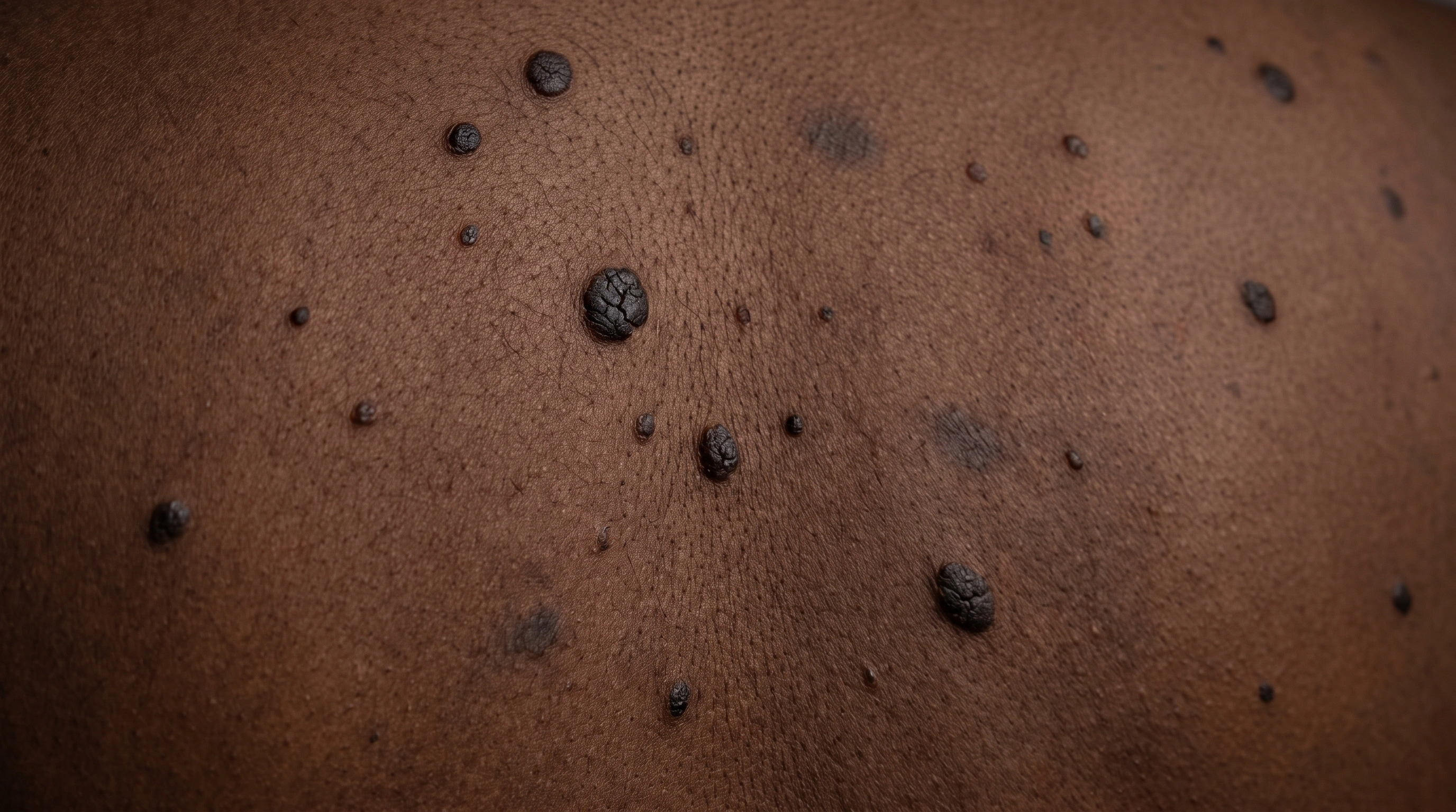
A cluster of black raised growths along the side of the neck on deep brown skin, each with a sharply defined edge, with two flat darker marks nearby where earlier growths were treated.
Diagram
Melanoma mechanism diagram. Cells multiply unevenly: The patch can become irregular. Pigment cells acquire DNA changes: Abnormal melanocytes escape normal growth control. Some tumors invade more deeply: Depth helps guide staging and treatment.
Seborrheic Keratosis mechanism diagram. A stuck-on plaque appears: The surface can look waxy or rough. Keratin collects in the growth: Tiny keratin-filled spaces may form. Surface cells grow locally: A benign epidermal growth develops.
In the skin

Abnormal pigment cells escape normal growth controls and multiply within the skin. Some melanomas grow into deeper tissue, and the depth of invasion helps determine their stage.

A benign overgrowth of surface skin cells creates a localized plaque. Keratin and sometimes pigment collect in the growth, giving it a waxy, rough or stuck-on appearance.

Where it shows up

Any skin site
Variable sites, often trunk or face

When it usually starts

010203040506070+

Melanoma. More common in adults, but a concerning changing lesion needs assessment at any age.

Seborrheic Keratosis. Usually appears in adult life and becomes increasingly common with age.

Causes and triggers

Causes

Melanoma starts when pigment cells develop DNA damage and grow out of control. Ultraviolet light is the main cause of that damage for most melanomas.

Seborrheic keratoses are harmless growths of skin cells that become more common with age.

Family tendency

About 1 in 10 people with melanoma have a family history of it. Genes such as CDKN2A raise the risk.

They often run in families.

Risk factors
Fair skin and red or blond hair, Many moles, Past blistering sunburns, Weakened immunity, Family history
Age over 50, Family history, Sun exposure
Trigger
● Melanoma
● Seborrheic Keratosis
Sunburns
Well established
Not listed
Tanning beds
Well established
Not listed
Intense, on-and-off sun
Strong link
Not listed
Aging
Not listed
Well established
Sun exposure
Not listed
Possible

Daily Care

Melanoma
Seborrheic Keratosis
Daily care

Habits that help

  • Learning the ABCDE signs
    Asymmetry, Border, Color, Diameter, Evolving above all.
  • The ugly duckling rule
    The mole that does not belong is worth examining.
  • Knowing ABCDE is not complete
    Firm, growing and new counts on its own.
  • Checking the places nobody checks
    Scalp, ears, soles, between the toes, nails, genitals and mouth.
  • Photographing your own skin
    Same distance and light, every few months.
  • Daily sun protection, no tanning beds
    Cuts future risk, and you control it.
  • Review immune-suppressing medicines with the prescriber; do not stop them yourself
    Immune suppression can change skin-cancer risk and follow-up needs. Do not stop a prescribed medicine without the specialist’s plan.

Avoid

  • Avoid: Tanning beds
    A recognized cause, riskier the younger you start.
  • Avoid: Blistering sunburns in childhood
    Linked more strongly than steady daily exposure.
  • Avoid: Only checking sun-exposed areas
    The scalp, soles, nails and mouth are found late.
  • Avoid: Assuming a pink bump is harmless
    Melanoma without pigment is a recognized variant.
  • Avoid: A fast-growing bump ignored
    Nodular melanoma grows downward from the start.
  • Avoid: Wearing sunscreen only on holiday
    Risk builds over a lifetime of ordinary days.
  • Avoid: Waiting out a changing spot
    A melanoma watched for six months is thicker.
  • Avoid: Treating a spot at home
    Freezing kits and black salve destroy the evidence.
  • Avoid: Skipping follow-up
    A second melanoma is meaningfully more likely.
  • Avoid: Not telling close relatives
    It changes their screening advice.
  • Avoid: Cryotherapy or cosmetic laser
    Freezing or lasering a pigmented spot off destroys it without anyone ever finding out what it was. If it was a melanoma, the thickness can no longer be measured and no one can check the edges. A spot that concerns you is examined and sampled, not erased for convenience, and this is one of the few places where the cheaper, quicker option costs something real.

Habits that help

  • Watch for change, not for existence
    Growing, darkening, bleeding or newly itchy.
  • Then leave it alone
    Avoid picking or rubbing a growth confirmed as benign.
  • Curettage or shave removal
    Precise, and it yields tissue to examine.
  • Daily sunscreen on treated areas
    Keeps the mark from deepening.
  • Discuss how age affects monitoring and recovery
    The biggest driver of how many you get.
  • Tell the clinician about your family history
    Why one person has one growth and another fifty.

Avoid

  • Avoid: Sun exposure
    Adds to the count, and darkens marks after removal.
  • Avoid: Assuming a dark growth is benign
    Melanoma is mistaken for one.
  • Avoid: Picking or peeling one off
    They bleed, inflame, and mark.
  • Avoid: Friction from clothing
    Straps, waistbands and belts irritate growths.
  • Avoid: Shaving over them
    The usual cause of sudden bleeding.
  • Avoid: Home freezing kits and acid pens
    They scar something nobody examined.
  • Avoid: A sudden crop of many new ones
    Many at once should be assessed.
  • Avoid: Removal without a diagnosis
    Destroyed tissue cannot be analyzed.
  • Avoid: Home freezing and acid kits
    Do not freeze or burn unexamined growths at home; tissue may need examination.

Treatment differences

Evidence labels come from each condition’s own treatment list.

Treatment
● Melanoma
● Seborrheic Keratosis
Over-the-counter
None listed
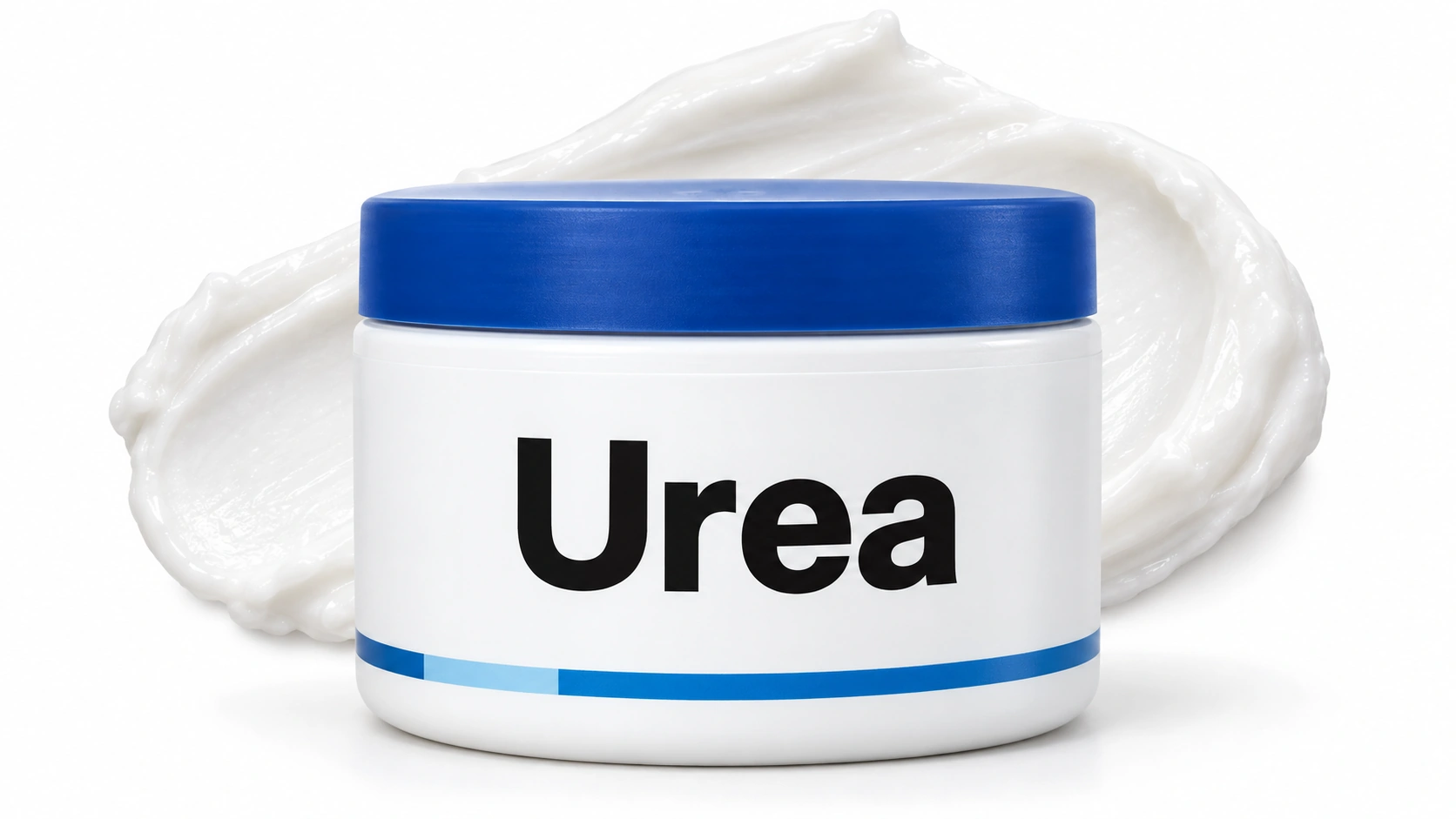
Five white jars of 40% urea cream lined up, several also listing 2% salicylic acid

Urea

Limited evidence

A strong urea cream can soften and flatten the rough white growths that scatter over the lower legs, called stucco keratoses, if the roughness is what bothers you. It smooths the surface rather than removing the growth, and it has to be used continuously. It does nothing for thick, dark, stuck-on growths elsewhere.

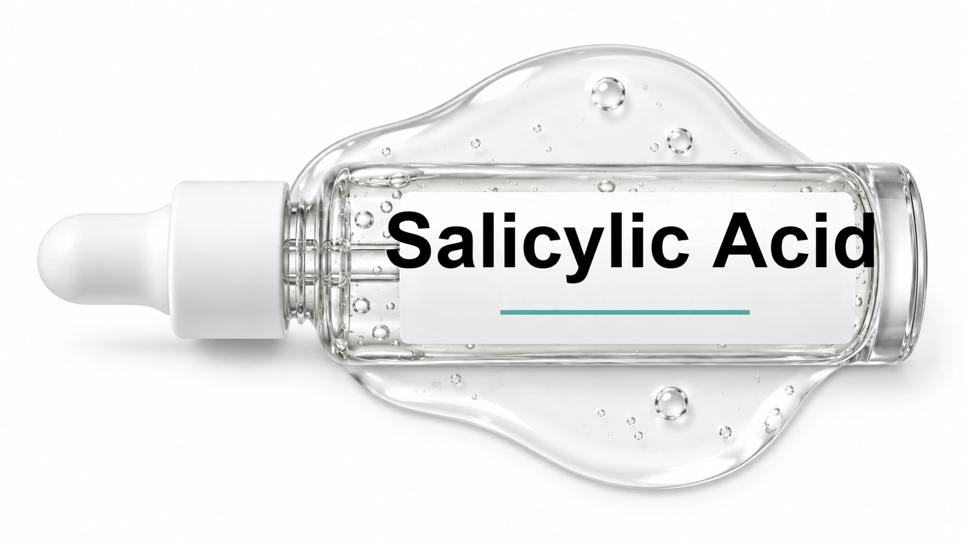
Paula's Choice 2% BHA exfoliant, La Roche-Posay Effaclar cleanser and a CeraVe psoriasis cream pump

Salicylic Acid

Limited evidence

Wart treatments are often tried on these because the surface looks similar. Salicylic acid can thin the rough top layer, but it does not clear the growth, and it irritates the normal skin around it. The bigger problem is that it is usually being used on something nobody has examined.

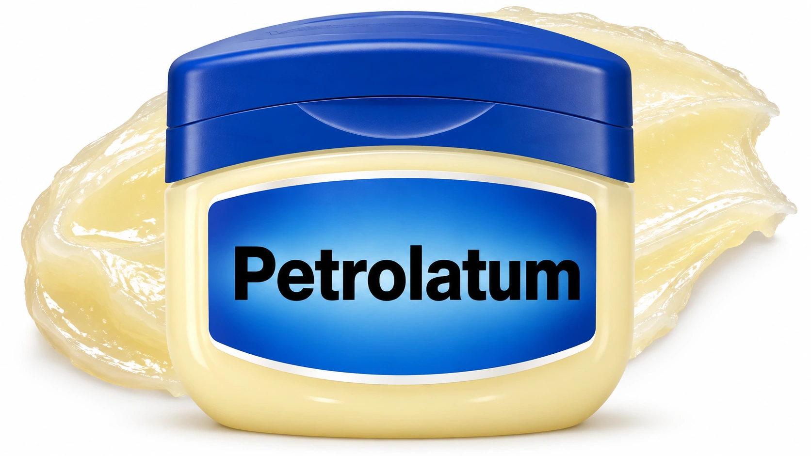
Four ointments on white: CeraVe Healing Ointment, a Vaseline jar, a petroleum jelly tube and Aquaphor

Petrolatum

Listed

This is aftercare, not treatment. A thin layer of plain petroleum jelly over a treated spot keeps the scab soft and gives the neatest healing, under a small dressing rather than an adhesive strip. It does nothing to a growth that is still there.

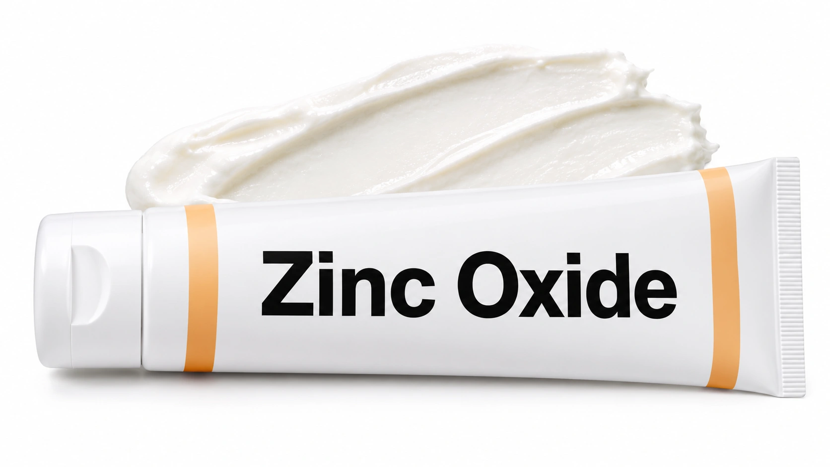
Four mineral sunscreens on white: Native SPF 30, Thinksport SPF 50, Colorescience Flex and EltaMD UV Clear

Zinc Oxide

Listed

Daily mineral sunscreen over a spot that has just been treated stops the temporary mark from darkening and helps it fade faster, which matters most on the face, chest and hands. It will not remove a growth or stop new ones forming. Sun is only one of several things that drive these, and age and family history matter more.

Prescription topicals
None listed

Eskata

Moderate evidence

A high-strength peroxide solution applied in clinic was approved for raised growths on the face and body. It cleared some growths, often needed repeat visits, and commonly caused stinging, crusting and pigment change. It was withdrawn from sale in the United States, so in practice removal is done by hand rather than with a liquid.

Procedures
Editorial illustration for Surgical Excision, based on the entry’s treatment or equipment reference.

Surgical Excision

Strong evidence

The treatment that cures the great majority of thin melanomas. Once the diagnosis is confirmed, a second and wider operation removes a margin of surrounding skin, with the width set by how thick the melanoma was. The scar is longer than most people expect, and that is the trade that stops it coming back at the site. Anything pigmented that is removed, anywhere, should be sent for pathology.

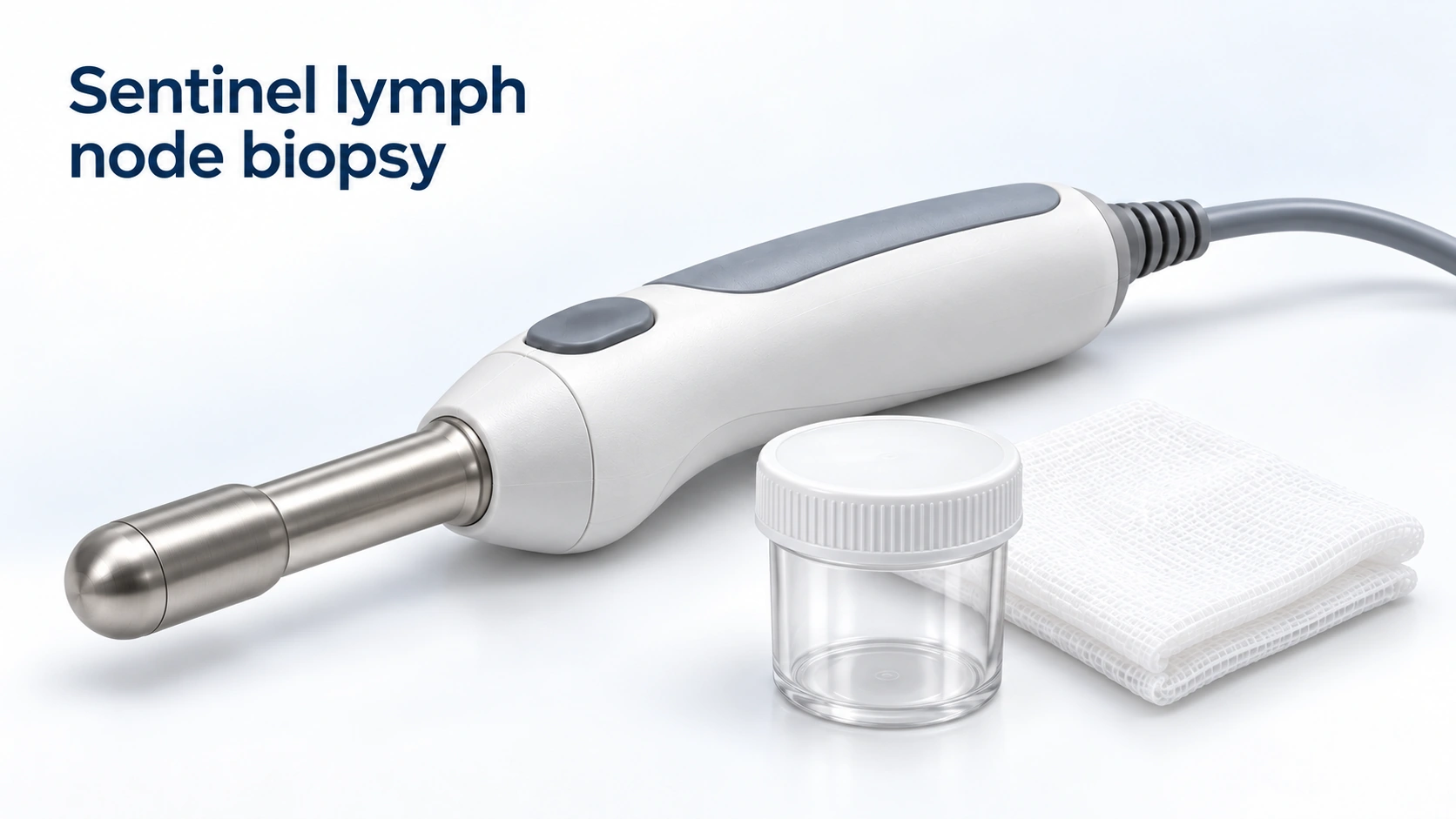
Editorial illustration for Sentinel lymph node biopsy, based on the entry’s treatment or equipment reference.

Sentinel lymph node biopsy

Strong evidence

Above a certain thickness, the first lymph node the area drains to is located with a tracer and removed to see whether cells have reached it. It is a staging test rather than a treatment: it tells you where you stand and shapes what is offered next. It carries its own risks, including lasting swelling of the limb, so it is offered as a discussion rather than an automatic step.

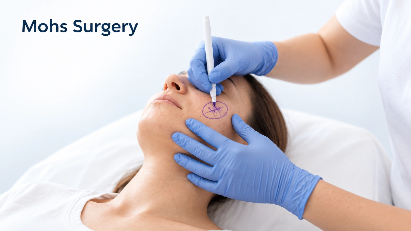
Editorial illustration for Mohs Surgery, based on the entry’s treatment or equipment reference.

Mohs Surgery

Moderate evidence

Used at some centers for lentigo maligna on the face, where sparing skin matters and the true edge of the pigment is hard to see. It is not the standard operation for melanoma generally — wide excision is, and the evidence sits with that. If Mohs is offered to you, it is fair to ask why it suits this particular lesion in this particular place.

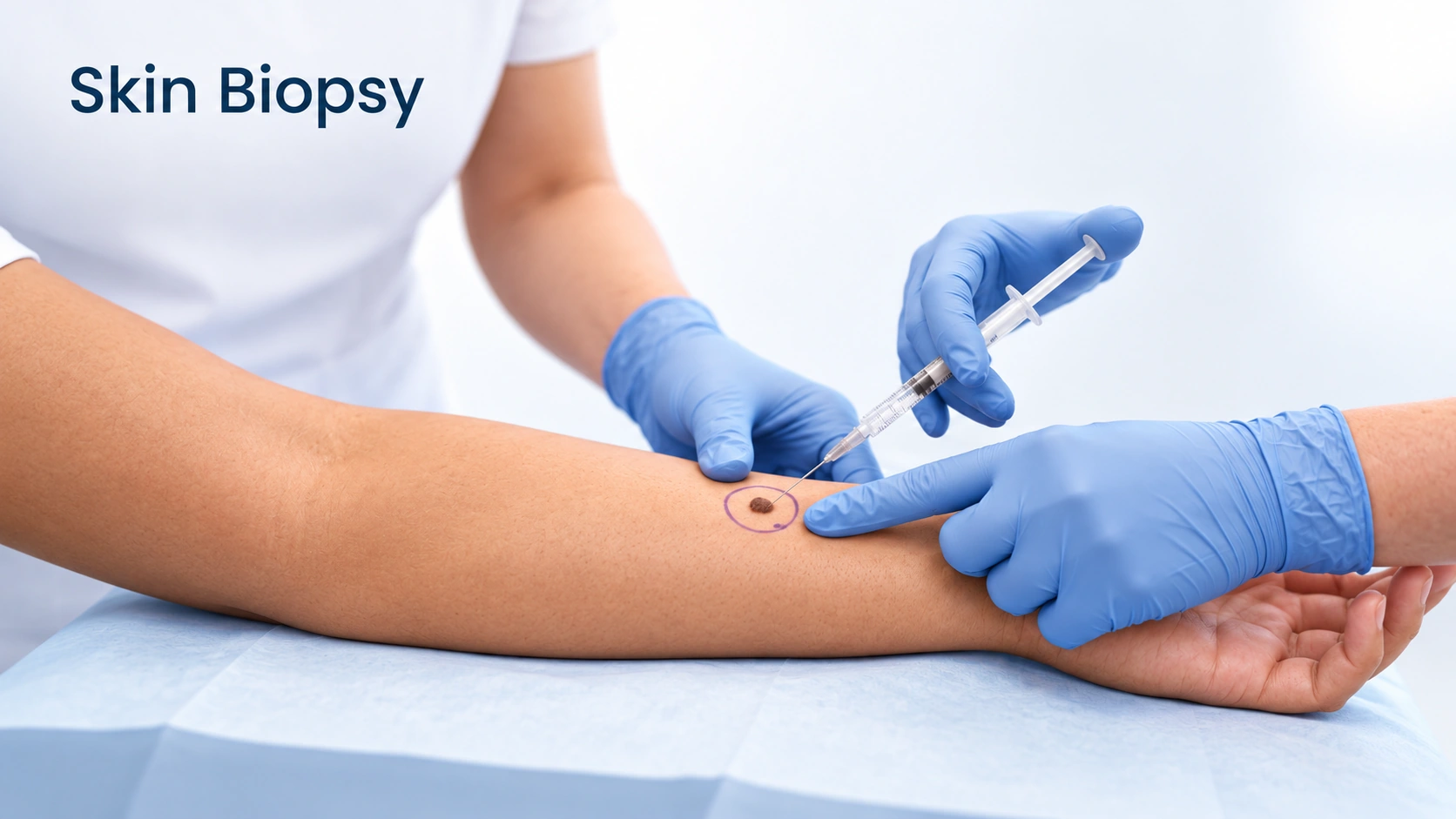
Editorial illustration for Skin Biopsy, based on the entry’s treatment or equipment reference.

Skin Biopsy

Listed

Nothing diagnoses melanoma except a biopsy. Where it is possible, the whole spot is removed with a narrow margin rather than sampled, because the pathologist needs the full depth to measure the thickness, and thickness sets everything that follows. It is done under local anesthetic and usually takes under half an hour. A biopsy does not make melanoma spread, and believing that it does is a common cause of delay.

Editorial illustration for Skin Biopsy, based on the entry’s treatment or equipment reference.

Skin Biopsy

Listed

When a growth is changing, bleeding on its own, or simply looks unlike your others, part or all of it is taken after numbing and examined under a microscope. It is the only way to be certain, and the threshold for doing it is deliberately low, because the cost of a small biopsy is trivial next to missing a melanoma. An irritated growth is one of the commonest reasons a benign growth ends up being sampled.

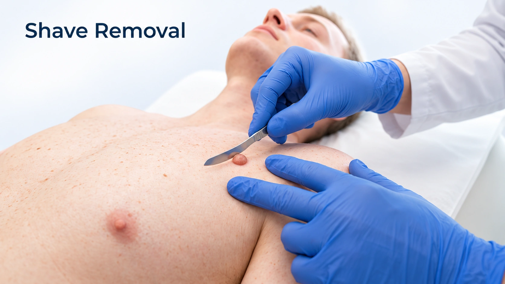
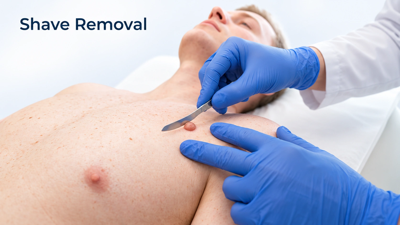
Editorial illustration for Shave Removal, based on the entry’s treatment or equipment reference.

Shave Removal

Moderate evidence

The growth is numbed and shaved off level with the skin in a few seconds. It is the method of choice when there is any question about what a growth is, because it produces tissue that can be sent to the laboratory. It leaves a flat mark that fades over months, and it does not stop new growths appearing elsewhere.

Editorial illustration for Electrodesiccation and Curettage, based on the entry’s treatment or equipment reference.

Electrodesiccation and Curettage

Moderate evidence

The growth is scraped off with a small curette and the base sealed with a fine electric tip. It is controlled and shallow, which is why it is often preferred for small facial growths and for the dark facial growths common on deeper skin tones. Scraping first still gives tissue that can be examined if there is doubt.

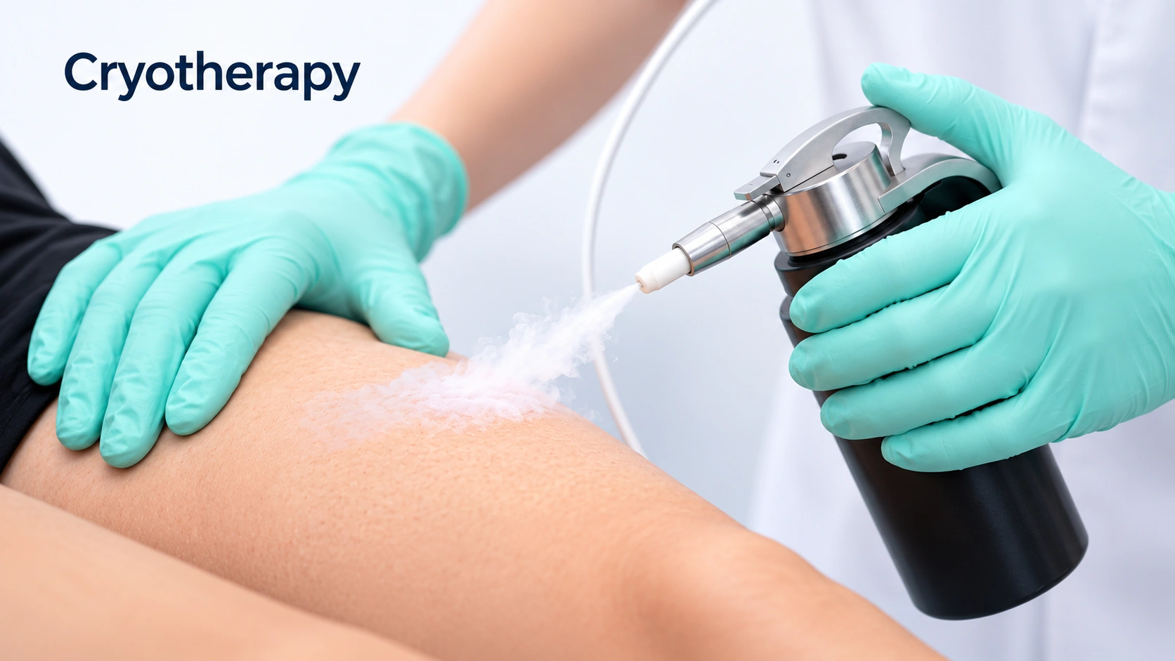
Editorial illustration for Cryotherapy, based on the entry’s treatment or equipment reference.

Cryotherapy

Moderate evidence

A few seconds of liquid nitrogen makes flatter growths blister and lift off over a week or two, and it is the quickest way to treat several at once. It destroys the growth, so nothing can be examined afterward, which is why it is not used on anything with a question attached. It is used cautiously on the face and on deeper skin tones, because the pigment cells are sensitive to cold and it can leave a pale spot.

Editorial illustration for Fractional CO2 Laser, based on the entry’s treatment or equipment reference.

Fractional CO2 Laser

Limited evidence

An ablative laser can vaporise a growth precisely and is sometimes used when several need treating on the face. Like freezing, it destroys the tissue, so it is only for growths already confirmed as harmless. It is the most expensive of the options and it is not better than a curette for a single growth.

Outlook

Long term

Melanoma does not progress on a fixed schedule. Some variants change over years, others over weeks, so this is the sequence, not a timetable. Skin checks continue for years after treatment, because a second melanoma and a recurrence are both possible, and close relatives are usually advised to have their skin checked.

Seborrheic keratoses appear slowly, change slowly, then stay. Two departures deserve attention: one growth that starts changing - growing, darkening, bleeding on its own, or itching persistently - and a sudden crop of many new ones over weeks to months.

Complications

Possible complications
  • Cells travel to the nearest nodes first
  • Treated by an oncology team
More details

Cells travel to the nearest nodes first
A lump near a previous melanoma site is significant and checked promptly.

Treated by an oncology team
Melanoma can spread to organs far from the skin. Immunotherapy and targeted therapy have changed outcomes substantially in the last decade.

  • Catching on clothing makes it flare
More details

Catching on clothing makes it flare
A growth caught by clothing, a strap or a waistband can turn red, swollen, itchy, crusted or sore, and can bleed. That is an irritated seborrheic keratosis. It settles, but it looks alarming and often prompts removal or a sample.

When to get help

When to get help

Arrange prompt assessment of a new or changing unusual spot, especially an evolving lesion or one that bleeds. Diagnosis requires clinical evaluation and usually biopsy.

Have a new, changing, bleeding or uncertain growth assessed; skin cancer can sometimes resemble a harmless stuck-on growth.

Common questions

Melanoma:How quickly does melanoma grow?

It varies more than people expect. The common superficial spreading variant often changes over months to a few years. Nodular melanoma can grow downward over weeks to a few months. You cannot tell which you have by looking, so a changing spot gets examined now, not in six months.

Melanoma:Does having a mole removed spread cancer?

No, and this belief causes delay. Removing a suspicious lesion is how melanoma is diagnosed and how the thickness is measured, and thickness determines the treatment. There is no evidence a biopsy spreads it.

Melanoma:Should I have all my moles removed to be safe?

No. Most melanomas arise as new spots, so removing moles does not remove the risk and means many scars for no benefit. Many or unusual moles are a reason for regular examinations, not preventive removal.

Melanoma:I have a dark line on my nail. Is that melanoma?

Usually not. Pigmented nail bands are common and often normal in deeper skin tones, frequently on several nails at once. It needs examining if it is on one nail only, if it is getting wider, if it has more than one shade, if the nail is splitting, or if the pigment spreads onto the skin around it.

Melanoma:If it is removed and the margins are clear, am I finished?

The melanoma is treated, but you are not discharged. Having had one raises the chance of a second, so skin checks continue for years, and close relatives are usually advised to have their skin examined. Ask how often yours should be and who arranges it.

Melanoma:I never sunbathe. Do I still need to worry about a changing spot?

Yes. Sun exposure raises risk, but melanoma also occurs on soles, palms, under nails and on skin never exposed, and in people who have never burned. Sun history changes your risk profile, not whether a particular spot should be looked at.

Seborrheic Keratosis:Could this be melanoma?

It could be, which is why it should be looked at rather than diagnosed at home. Melanoma is sometimes mistaken for a seborrheic keratosis, and a dermatoscope resolves most of these in seconds. The concerning features are change in size, shape or color, bleeding without being knocked, an irregular edge, and a growth unlike your others. Book rather than watch.

Seborrheic Keratosis:Why am I suddenly getting so many?

Age and family history account for nearly all of it. They accumulate steadily from the forties onward, and someone whose parents had many will develop many. A genuinely sudden crop over weeks to months, especially with itching, is different and worth examining.

Seborrheic Keratosis:Does removing one stop more from appearing?

No. Removal deals with that growth, not the age and inherited tendency behind it, so new ones can keep appearing elsewhere. That is expected, not a failed removal.

Seborrheic Keratosis:Will removal leave a mark?

Often, at least for a while. A flat spot darker or paler than nearby skin is common, and dark marks usually fade over months. Pale marks can be permanent and are more noticeable on deeper skin tones, so ask about the method and do a test spot before treating a large area. Daily sunscreen on the treated spot makes a real difference.

Seborrheic Keratosis:Mine got red and itchy and started bleeding. What happened?

Most likely it caught on clothing, a strap or a razor and became an irritated seborrheic keratosis. They look far worse than they are and they settle. Still have it looked at - an irritated growth is one of the harder ones to be sure about by appearance, and a growth that bled with nothing catching it is a different question.

Seborrheic Keratosis:Can I have them all removed?

As many as you and your doctor think sensible, usually in sessions rather than all at once. It is normally considered cosmetic, so ask about cost first. On the face, and particularly on deeper skin tones, start with a small test area and see how it heals.
Sources

Sources and further reading

  1. DermNet
  2. DermNet
Dermapedia on YouTube