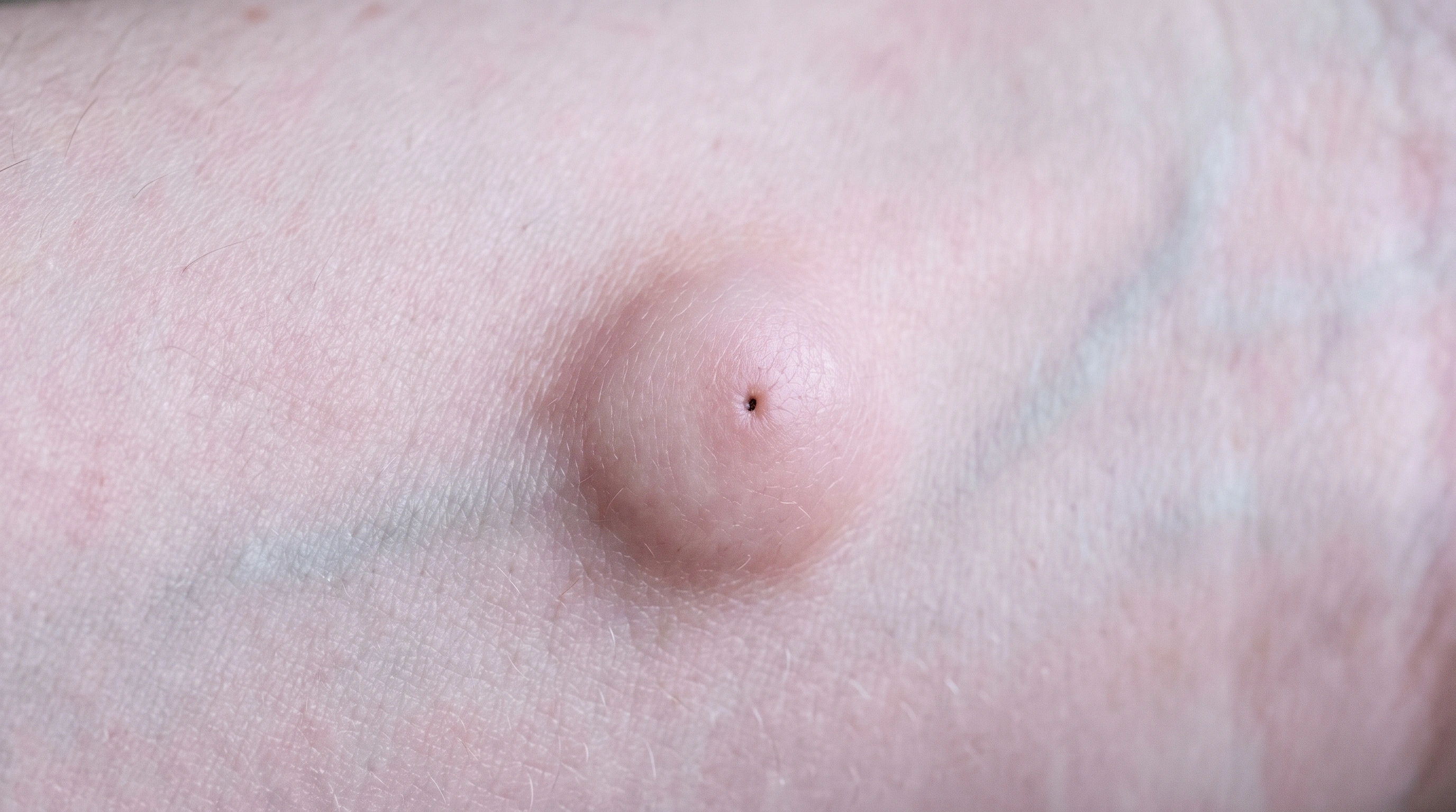
Dermatofibroma vs Moles

A common benign fibrous skin nodule. Moles are ordinary clusters of pigment cells in the skin, and almost all of them stay harmless for a whole lifetime. What matters is not how many you have, but whether one of them is changing - because a changing mole is the earliest sign of melanoma, and the earliest melanoma is the one that is easiest to cure.
Research updated 2026-10-23
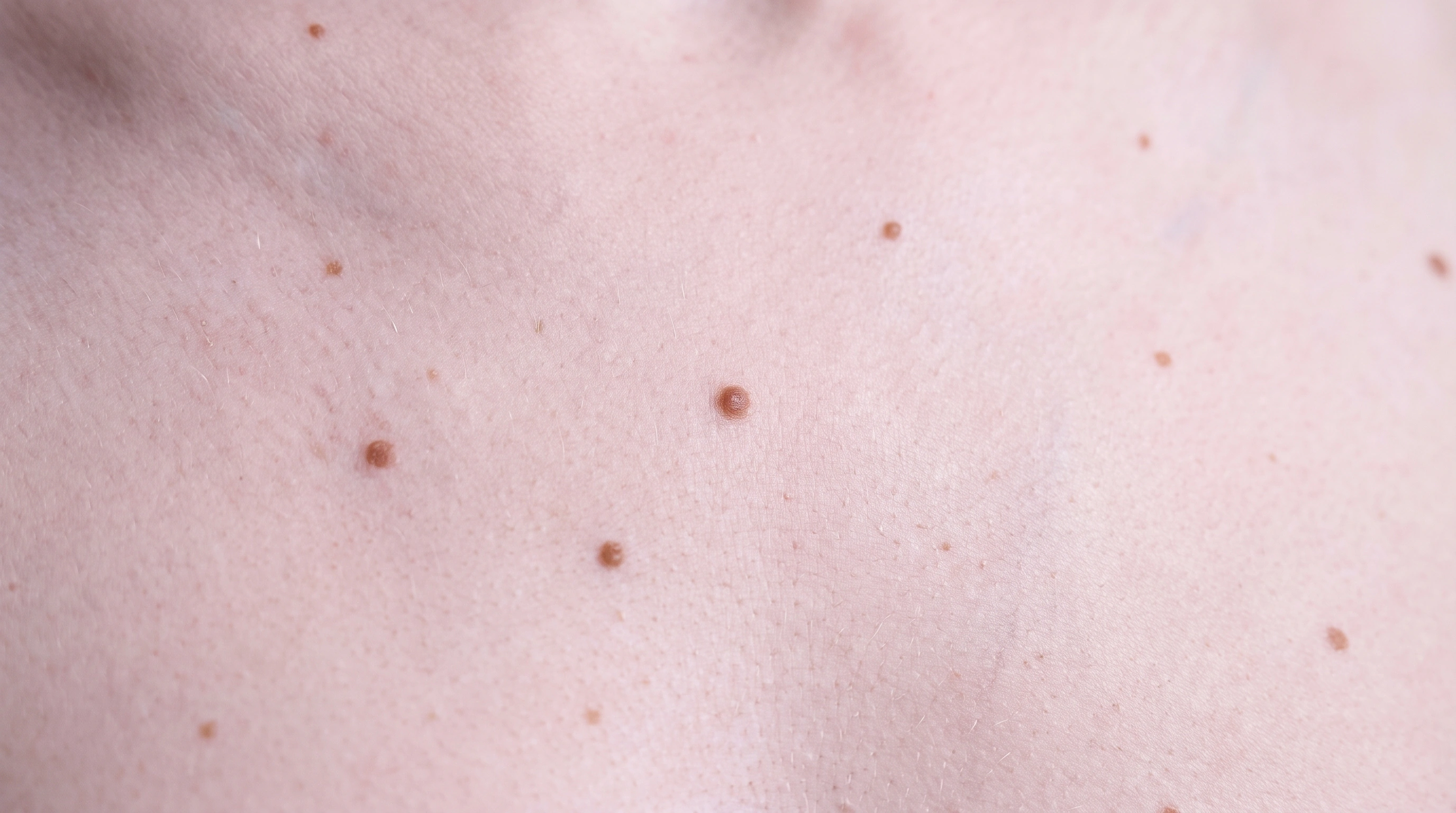
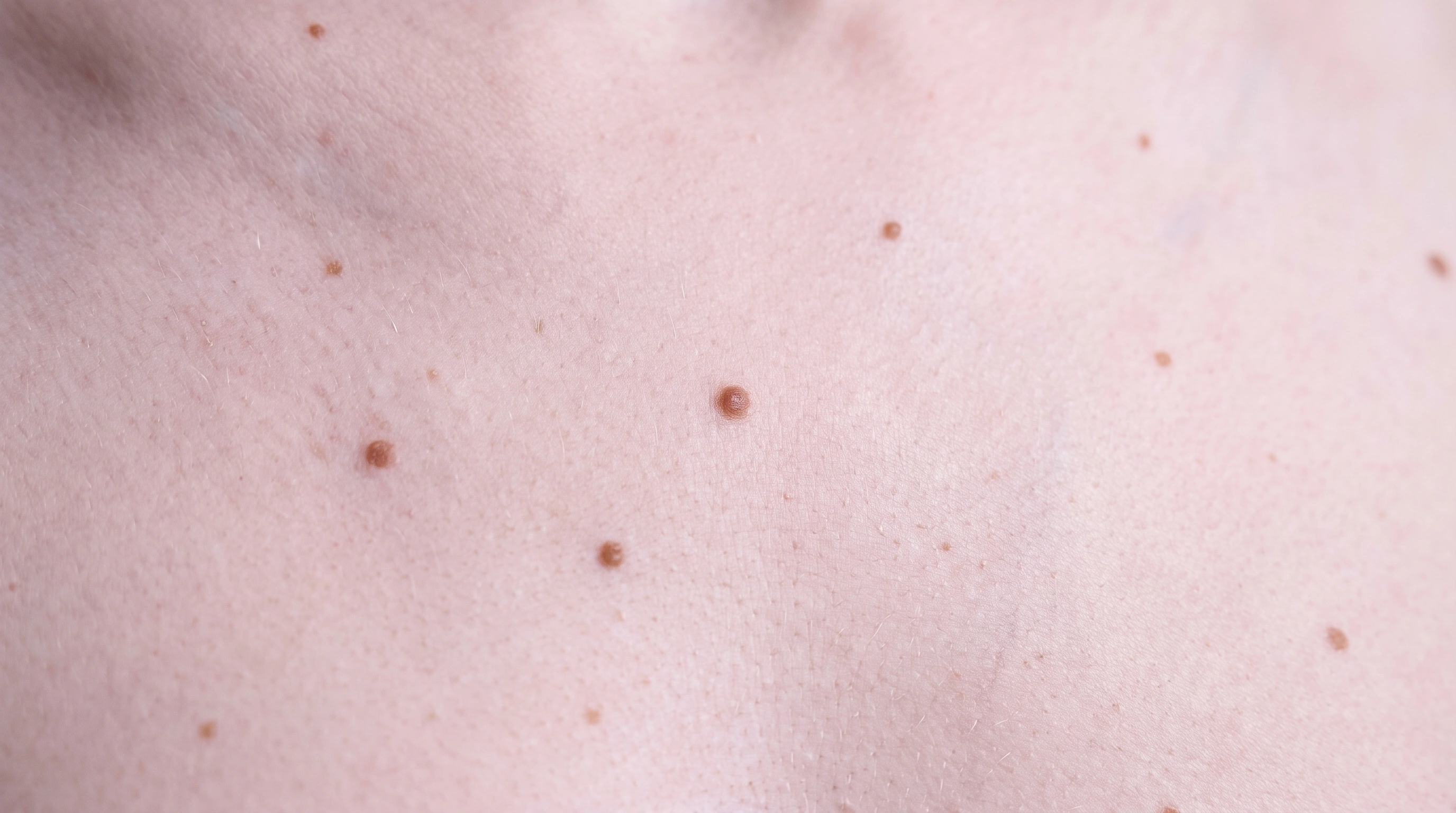
DermatofibromaSeveral small tan to mid-brown moles on the shoulder and upper arm of an adult with light skin, each evenly colored with smooth edges, with one larger mole among them.

Key differences

The same eight facts on every condition comparison.

Usual age
Adults
Childhood and teens
Where
Limbs, especially lower legs
Anywhere
Main sign
Firm bump that dimples when pinched
Even brown spots or bumps
Itch or pain
Sometimes itchy
No
Contagious
No
No
Long term
Harmless; stays
Harmless; watch for change
Scars
Removal leaves a scar
No
Who gets it
Adults, often after a bite or shave nick
Everyone; more in fair skin

What is it?

Dermatofibroma
Moles
What is it?
  • What it is
    A common benign fibrous skin nodule.
  • They are clusters of pigment cells.
    Moles are usually benign collections of melanocytes.
  • They vary in appearance.
    They can be flat or raised, skin-colored or pigmented.
  • They appear at different times.
    Some are present at birth; others appear later.
  • New or changing moles need a check.
    A stable familiar mole differs from an evolving lesion.

What it looks like

Dermatofibroma

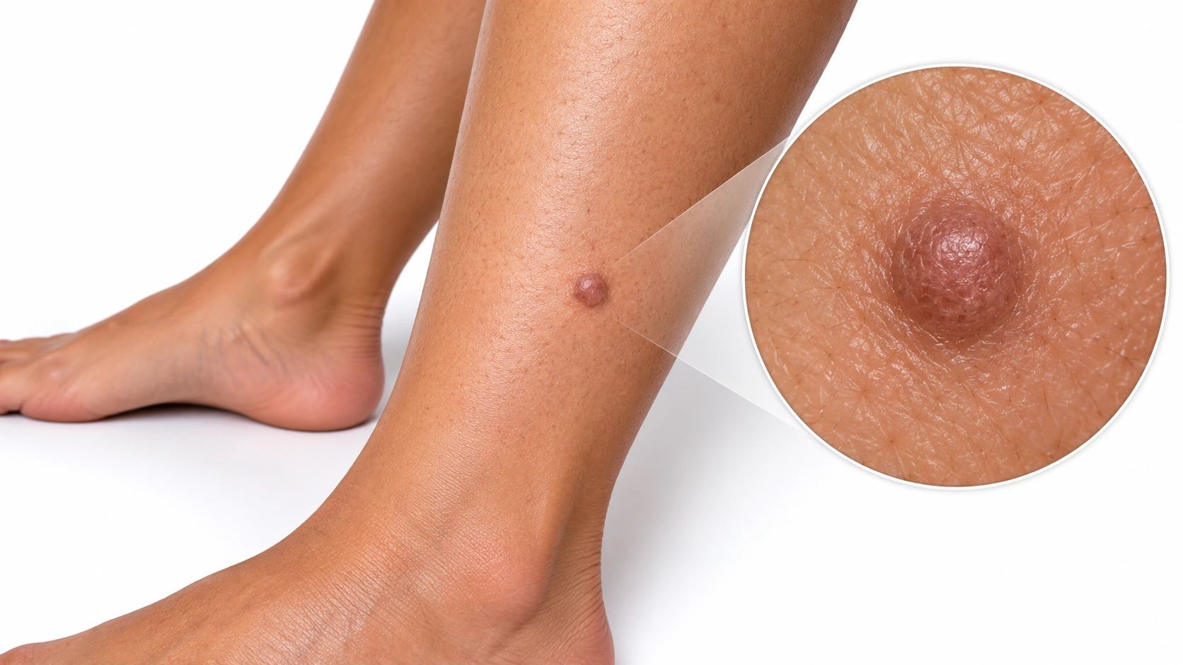
A lower leg with a small brown raised bump and a circular magnified detail.

Dermatofibroma An enlarged inset highlights the bump's shape and surface.
Moles

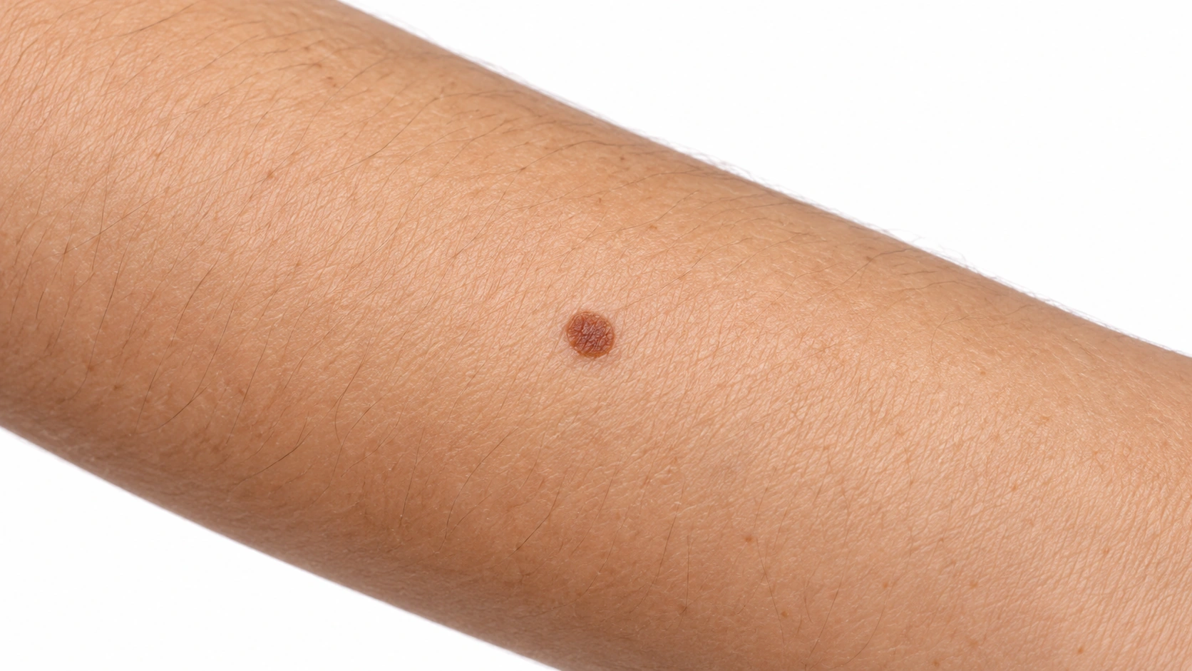
Common brown mole on the arm.

Common mole Small, round, even color.
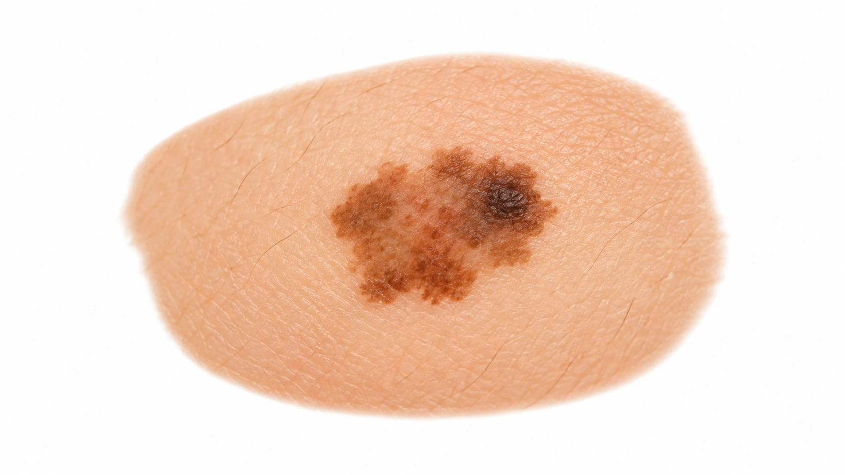
Representative image of irregular pigmented spot. It illustrates a visible feature or experience and is not a diagnostic reference.

Atypical mole Larger, uneven border or color.
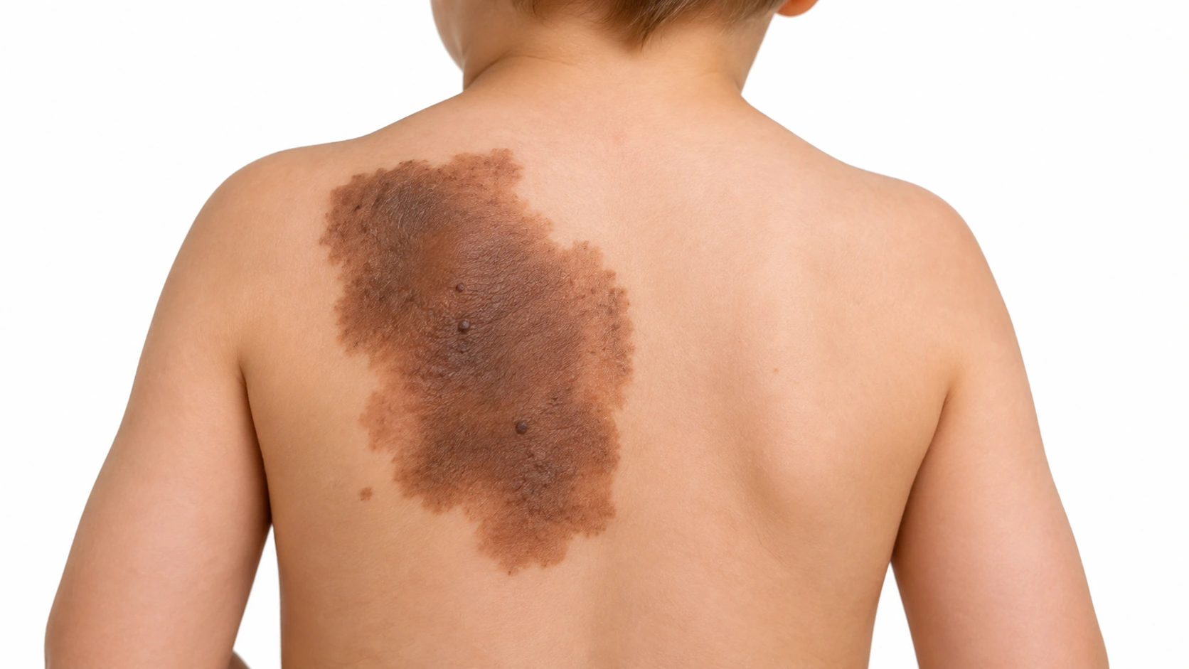
Large congenital mole on a child's back.

Congenital mole Present from birth.

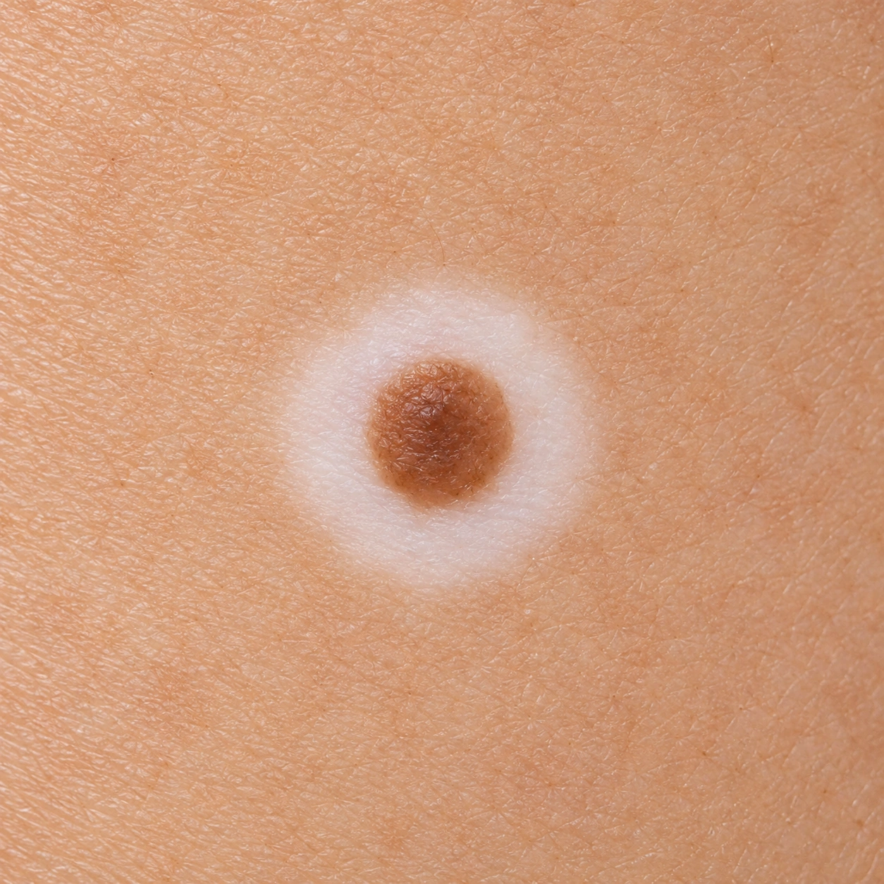
Generated image: Mole surrounded by a white ring of skin. Not a patient photograph.

Halo nevus Generated illustration. Mole surrounded by a pale ring.
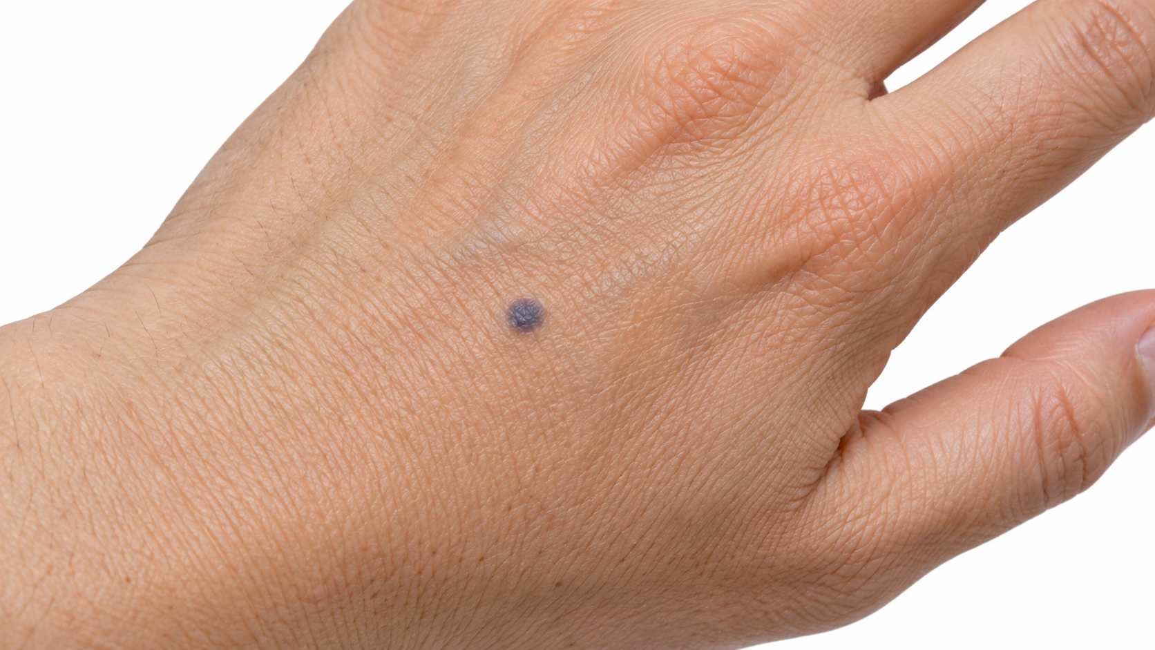
Small blue-gray mole on the hand.

Blue nevus Small blue-gray mole.
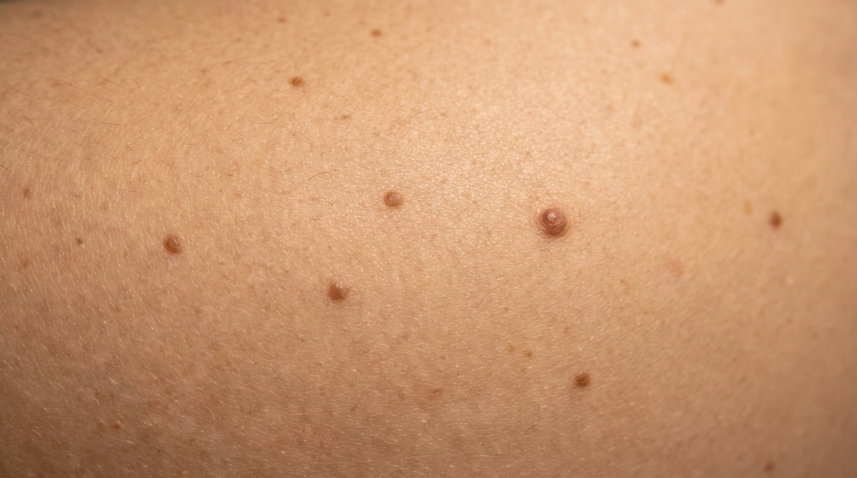
Skin tones
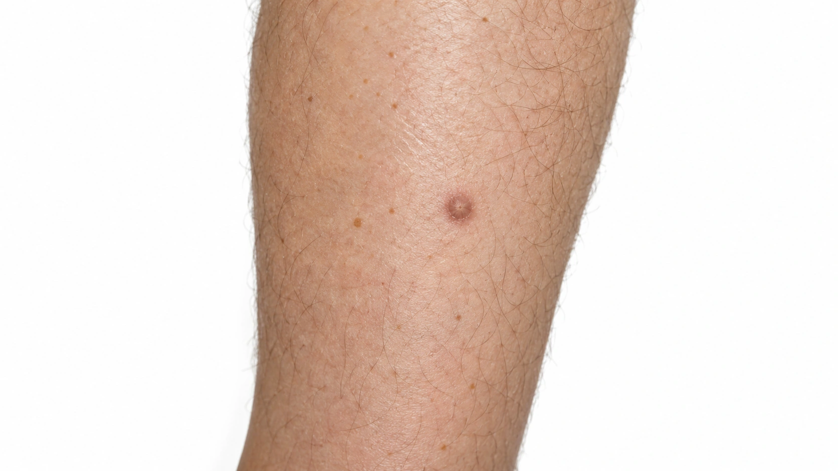
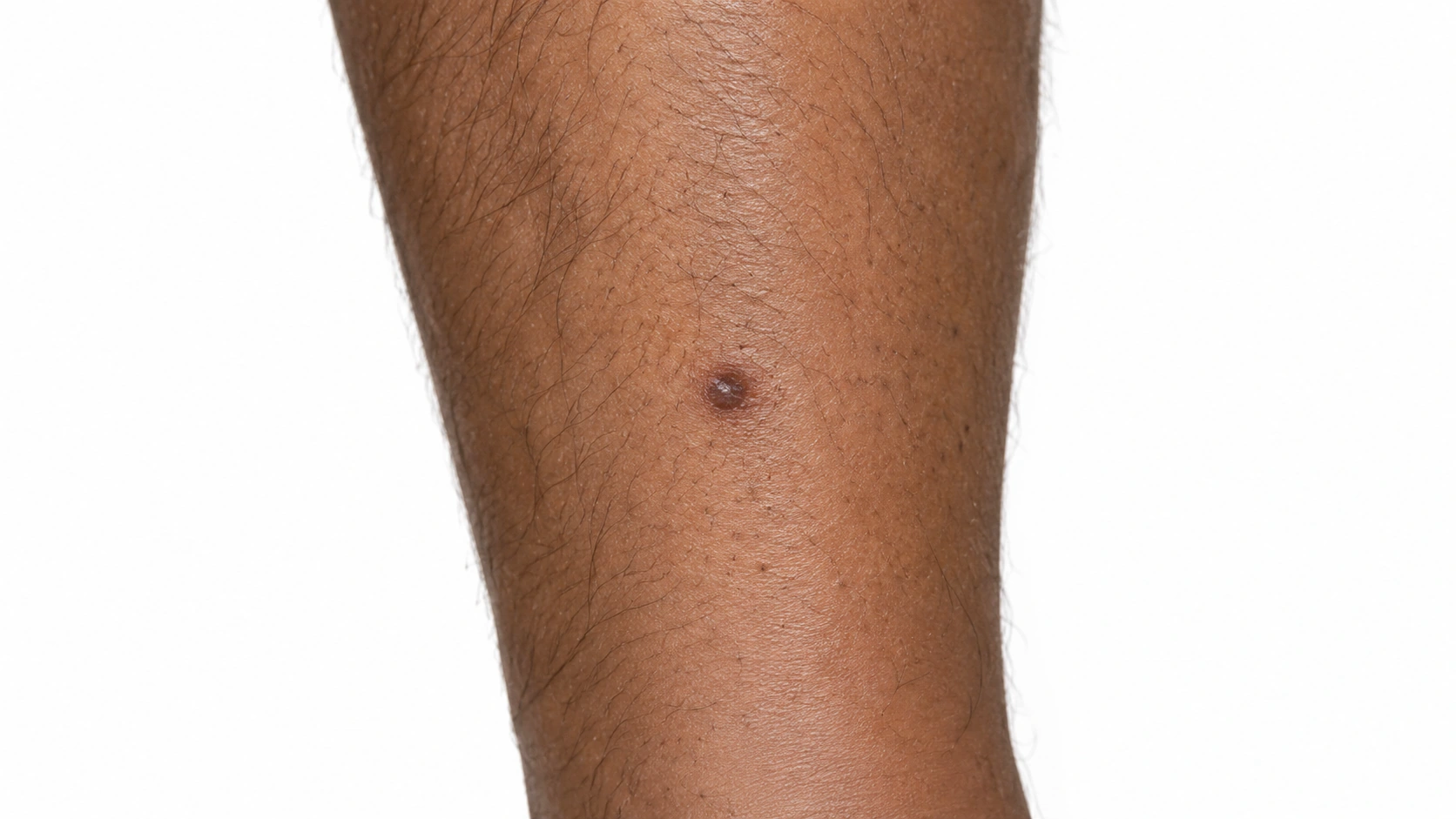
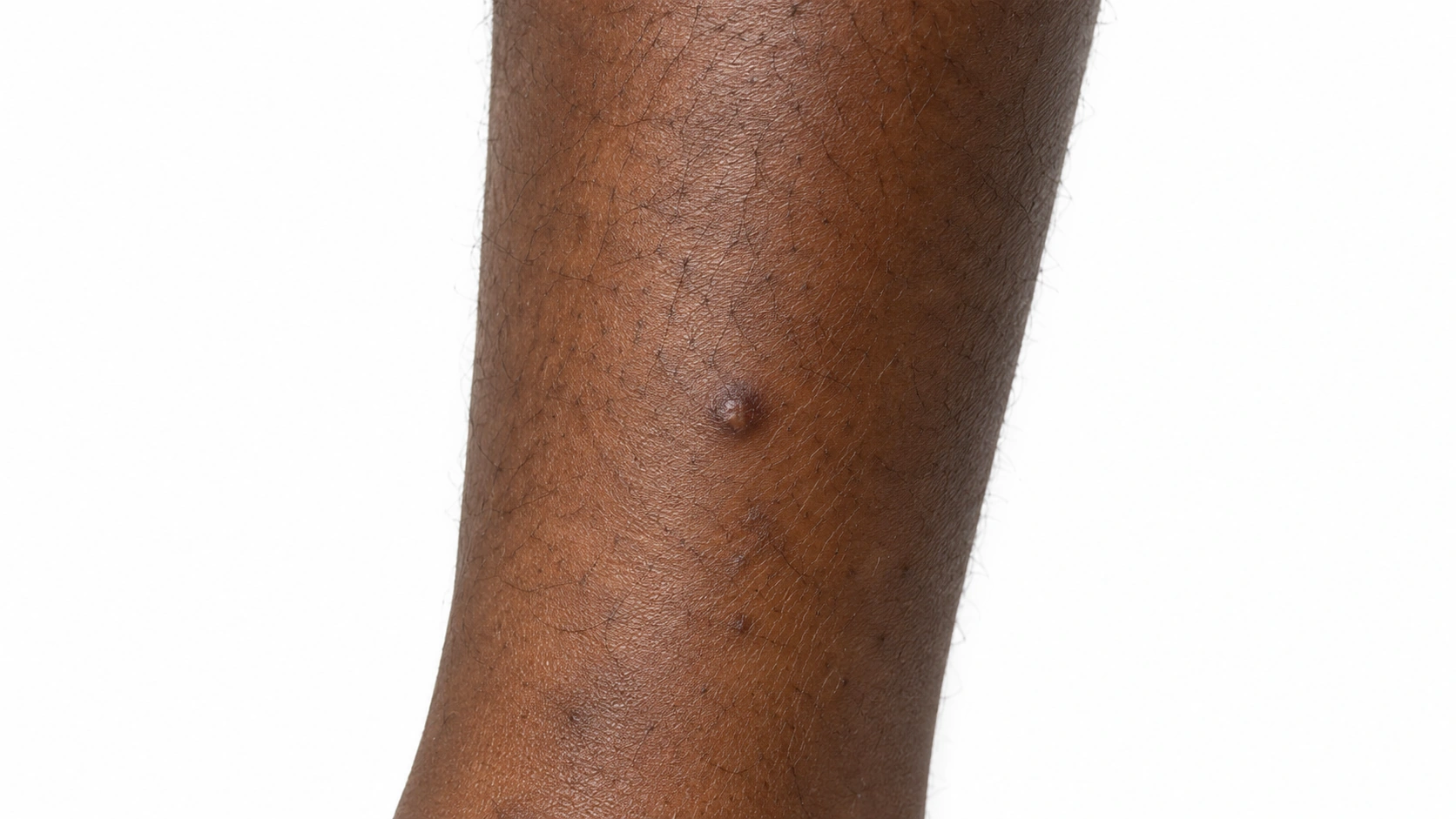
A small raised dermatofibroma on the lower leg.
Dermatofibroma on medium skin.
A small raised dermatofibroma on the lower leg.
Several small tan to mid-brown moles on the shoulder and upper arm of an adult with light skin, each evenly colored with smooth edges, with one larger mole among them.
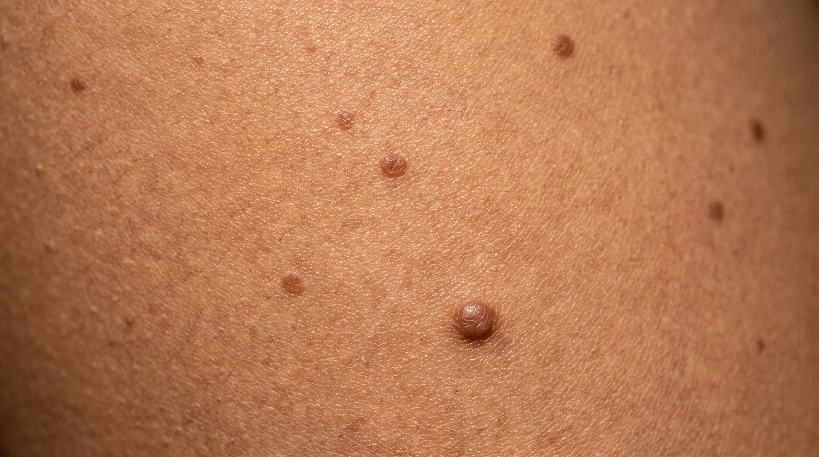
Several mid to dark brown moles on the shoulder and upper arm of an adult with medium olive-toned skin, with one larger mole that is uneven in shape.
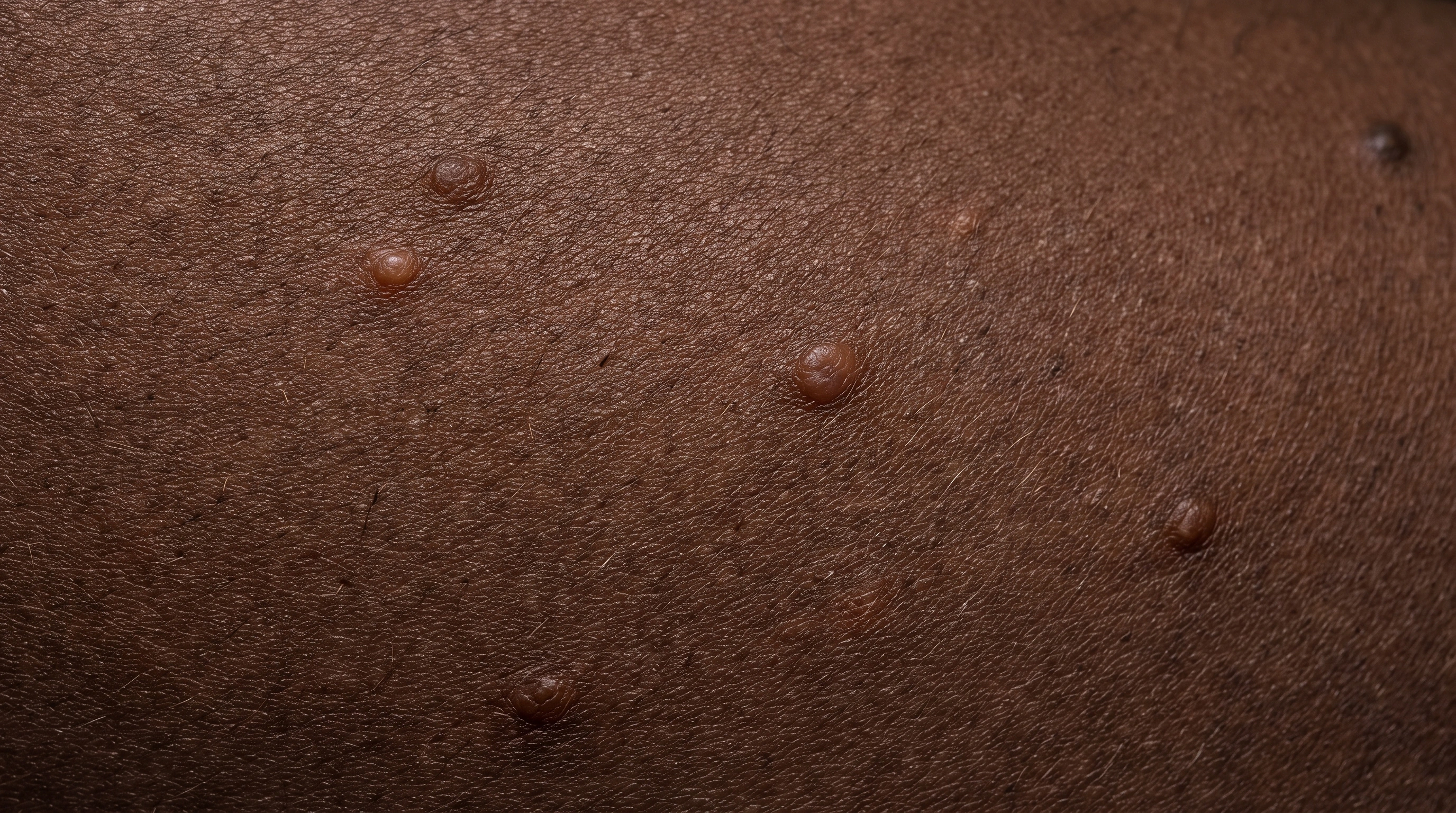
Several dark brown to near-black moles on the shoulder and upper arm of an adult with brown skin, each small, evenly colored and smooth-edged, on even-toned surrounding skin.
Several dark brown to near-black moles on the shoulder and upper arm of an adult with deep brown skin, each small, evenly colored and smooth-edged.
Diagram
Moles mechanism diagram. A spot or bump is visible: Many moles are flat, while others are raised. Pigment cells cluster: Nevus cells form small organized nests. Some nests sit in the dermis: Different mole types have different arrangements.
In the skin

A localized growth of fibrous cells forms a firm skin nodule. The reason the growth develops is often uncertain.

Moles form when benign pigment cells cluster into small nests. The location and arrangement of these cells help explain why some moles are flat and others are raised.

When it usually starts

010203040506070+

Dermatofibroma. Most often noticed in adults.

Moles. Most ordinary moles appear during childhood and adolescence; new or changing adult lesions deserve attention.

Causes and triggers

Causes

A local fibrous-cell proliferation forms a stable nodule; the cause is often uncertain.

Moles form when pigment cells grow in a cluster instead of spreading through the skin. Most appear in childhood and early adulthood.

Family tendency

The number of moles you have is largely inherited.

Risk factors
Adult age, Sometimes prior minor skin injury
Fair skin, Family history of many moles, Childhood sunburns
Trigger
● Dermatofibroma
● Moles
No established short-term flare trigger
Not a typical trigger
Not listed
Sun exposure in childhood
Not listed
Strong link
Pregnancy
Not listed
Possible

Daily Care

Dermatofibroma
Moles
Daily care

Habits that help

  • Leave a stable, clinician-confirmed benign lesion alone
    It may not need treatment if it is comfortable and not changing.
  • Reduce rubbing from clothing, shaving or hair tools
    Repeated injury can make a harmless lesion sore or cause bleeding.
  • Notice meaningful changes without repeatedly squeezing it
    Arrange examination for rapid growth, persistent bleeding or a change in appearance.

Avoid

  • Avoid: Cutting, burning, freezing or acid removal at home
    Home removal can cause infection, scarring and delay the correct diagnosis.

Habits that help

  • Learn the ABCDE signs
    Asymmetry, Border, Color, Diameter, Evolving — the one that matters most.
  • Learn the ugly duckling sign
    The mole that does not belong in the family.
  • Check your own skin about once a month
    Include back, scalp, soles, palms and nails.
  • Take photographs
    Comparison over months beats memory.
  • Use sunscreen daily on exposed skin
    Broad spectrum, SPF 30 or higher, reapplied outdoors.
  • Use shade, clothing and hats
    A long-sleeved shirt outperforms any bottle.
  • Never use a tanning bed
    No protective base tan, no safe dose.
  • Protect children from burning
    Risk is largely set before adulthood.
  • Discuss skin surveillance if your immune system is weakened
    Immune-suppressing treatment raises skin cancer risk.

Avoid

  • Avoid: Sunburn
    Raises mole count and damages the cells inside.
  • Avoid: Tanning beds
    A concentrated dose of the damaging wavelengths.
  • Avoid: Intense occasional sun
    Holiday exposure matters more than steady daily sun.
  • Avoid: Skipping sunscreen in cloud and winter
    Ultraviolet passes cloud and reflects off snow.
  • Avoid: Assuming a painless mole is fine
    Pain and itch are late signs.
  • Avoid: Only checking the parts you can see easily
    Scalp, back, buttocks, soles and nails all count.
  • Avoid: Relying on a phone app
    A reassuring result is no reason to cancel.
  • Avoid: Being told once that a mole is fine
    A mole examined five years ago was fine five years ago.
  • Avoid: A mole-checking phone app
    Use photos to track changes; never delay an examination because an app gives reassurance.
  • Avoid: Mole removal creams and kits
    Do not burn, freeze or dissolve moles at home; the tissue may need examination.
  • Avoid: Waiting to see if a change settles down
    Three months of waiting is three months of growth.
  • Avoid: Cryotherapy
    Freezing is excellent for several other skin growths and is the wrong tool for a mole. It destroys the tissue, so nothing can be examined, and it commonly leaves the deeper pigment cells behind along with a pale mark. Lasering or burning a mole off has the same two problems. If a mole is worth removing, it is worth removing in a way that can be checked.

Treatment differences

Evidence labels come from each condition’s own treatment list.

Treatment
● Dermatofibroma
● Moles
Procedures

Excision if indicated

Listed

Pain, growth or diagnostic uncertainty may justify removal or tissue assessment.

Editorial illustration for Surgical Excision, based on the entry’s treatment or equipment reference.

Surgical Excision

Strong evidence

The standard for any mole that is genuinely in doubt: the whole mole is cut out under local anesthetic with a narrow margin of normal skin and sent for examination under a microscope. Taking all of it matters, because the pathologist has to see the full depth and the edges. It leaves a line scar, and results come back in a week or two.
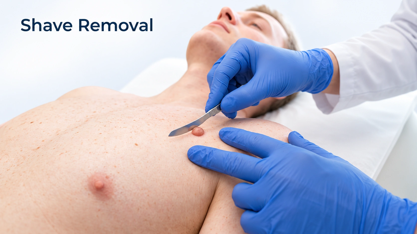
Editorial illustration for Shave Removal, based on the entry’s treatment or equipment reference.

Shave Removal

Moderate evidence

A raised mole that catches on a razor or a collar can be shaved off flush with the skin under local anesthetic, leaving a flat pale mark rather than a line scar. It is a reasonable choice for a mole that is not suspicious, and the tissue still goes to the laboratory. Two honest limits: pigment left at the base can grow back, and a shave through a melanoma cuts across it rather than under it.

Editorial illustration for Punch Biopsy, based on the entry’s treatment or equipment reference.

Punch Biopsy

Listed

A small circular blade removes a small mole whole, down through the full thickness of the skin, and it is closed with a stitch or two. It suits moles smaller than the punch. Used to take a sample from part of a larger suspicious mole it can under-read the depth, so for those an excision is the better choice.

Outlook

Long term

A confirmed dermatofibroma is harmless and often stays for years. Removal is optional when troublesome, leaves a scar and may not prevent recurrence if the lesion is incompletely removed.

Most moles remain harmless, but their appearance can change over a lifetime. A changing mole, a new unusual pigmented spot or one that looks different from the others needs examination. Monitoring should follow the person's individual risk.

Complications

Possible complications

A secure diagnosis matters because other lesions can look similar.

Most moles are benign. Having many or atypical moles can mark increased melanoma risk, and a changing lesion should be examined rather than assumed to be an ordinary mole.

When to get help

When to get help

Have a new, growing, bleeding, painful or otherwise changing lesion examined.

Arrange prompt assessment for an evolving, unusual or bleeding mole. Comparing a spot with your other moles is useful, but photographs cannot confirm a diagnosis.

Common questions

Dermatofibroma:What is Dermatofibroma?

A common benign fibrous skin nodule.

Dermatofibroma:What can I do day to day?

Avoid picking or home removal. Protect exposed skin from the sun.

Dermatofibroma:When should I get medical advice?

Have a new, growing, bleeding, painful or otherwise changing lesion examined.

Moles:How do I tell a normal mole from a dangerous one?

Use two checks together. ABCDE covers asymmetry, an uneven border, more than one color, a diameter above about 6 millimeters, and - most important - evolving, meaning changing. The ugly duckling check asks whether the mole looks like your others. A spot that fails either, or passes both but keeps changing, should be examined in person.

Moles:Is it normal to get new moles as an adult?

New moles keep appearing through the teens and twenties and become uncommon after about 40. Many new pigmented spots in older adults turn out to be seborrheic keratoses or sun spots. That is a reason to have a new spot identified, not to assume it is one of those.

Moles:Can I have a mole removed just because I do not like it?

Yes, and it is a reasonable request. What matters is how it is done — cut out under local anesthetic and sent to a laboratory. Lasering, freezing or burning leaves nothing to examine, so ask for the version that gets checked.

Moles:Does having a lot of moles mean I will get melanoma?

No. Your risk is higher than average and regular checks are worth doing, but most people with a high mole count never develop melanoma. The response is a schedule of skin exams, not worry.

Moles:Can melanoma appear where there was no mole?

Yes — most melanomas arise on skin that had no mole before. That is why a new spot that grows, or a sore that will not heal, deserves the same attention as a changing mole.

Moles:My mole changed during pregnancy. Is that expected?

Moles commonly darken slightly in pregnancy, and skin stretching over a growing abdomen or breasts can change a mole's shape. Melanoma does occur in pregnancy, though. A mole that changes in shape, border or color should be looked at while you are pregnant, not after.

Sources

Sources and further reading

  1. Dermatofibroma
  2. DermNet
Dermapedia on YouTube