Squamous Cell Carcinoma vs Warts

Squamous cell carcinoma is the second most common skin cancer. It usually appears as a rough, scaly or crusted bump on skin that has had years of sun, and it often looks like a sore that will not heal. Found early it is removed and cured. Left alone it grows deeper, and a small share spread. Warts are firm, rough growths caused by a common virus that infects the top layer of skin. They are harmless and most of them clear on their own, but they spread easily and they are slow to treat.

Reviewed by Dr. Schwarz, Board-Certified Dermatologist · Research updated 2026-10-23
Crusted red raised lesion on a light-skinned sun-exposed hand.A rough, dome-shaped wart on the side of a finger on fair skin. The bump is pinkish with a thin red ring at its base, and several tiny black dots are clearly visible in the thickened surface.

Key differences

The same eight facts on every condition comparison.

Usual age
50 and over
5 to 20
Where
Face, ears, scalp, hands
Hands, fingers, feet
Main sign
Scaly bump or sore that won't heal
Rough firm bump with black dots
Itch or pain
Can be tender
Usually painless
Contagious
No
Yes, mildly
Long term
Cured if removed early
Most clear in 1 to 2 years
Scars
After removal
No
Who gets it
Fair skin, sun exposure, immunosuppression
Children and teens

What is it?

Squamous Cell Carcinoma
Warts
What is it?

It is a skin cancer of keratinocytes.
It arises from the main cells of the outer skin.

It often looks crusted or ulcerated.
It may be a growing scaly lump, persistent sore or tender patch.

Most are treated successfully.
Some can invade deeply or spread to lymph nodes.

Risk is assessed individually.
Size, depth, site and immune status guide care.

They are caused by HPV.
Cutaneous warts are benign growths caused by certain types of human papillomavirus.

They spread by contact.
Warts can pass through direct contact or from one area of skin to another.

Many clear on their own.
Many resolve naturally, especially in children.

Diagnosis is usually visual.
Clinicians look for the rough surface and small capillary dots; uncertain growths may be biopsied.

What it looks like

Squamous Cell Carcinoma

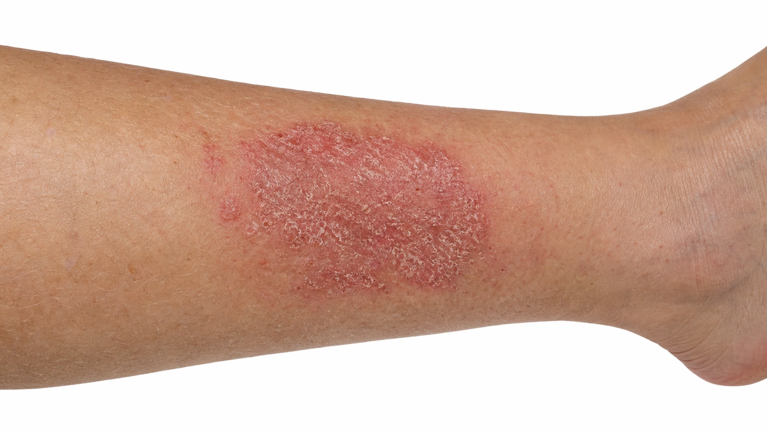
A sharply defined red scaly patch on the lower leg, representing squamous cell carcinoma in situ.

Squamous cell carcinoma in situ Flat red scaly patch, surface only.

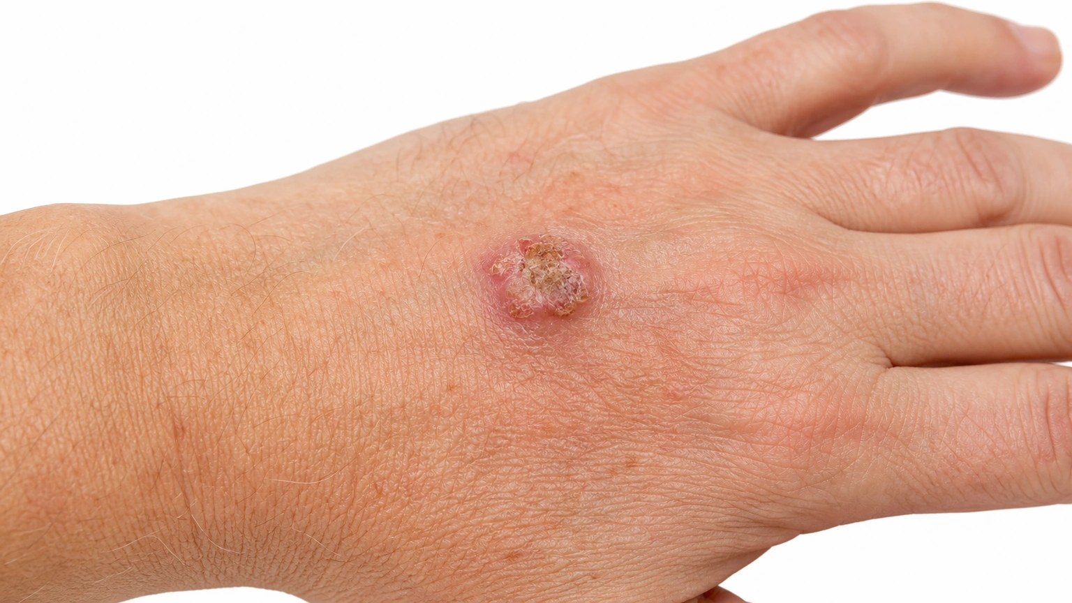
A scaly red raised bump on the back of the hand, representing squamous cell carcinoma.

Invasive squamous cell carcinoma Generated illustration. Firm, crusted or ulcerated bump.

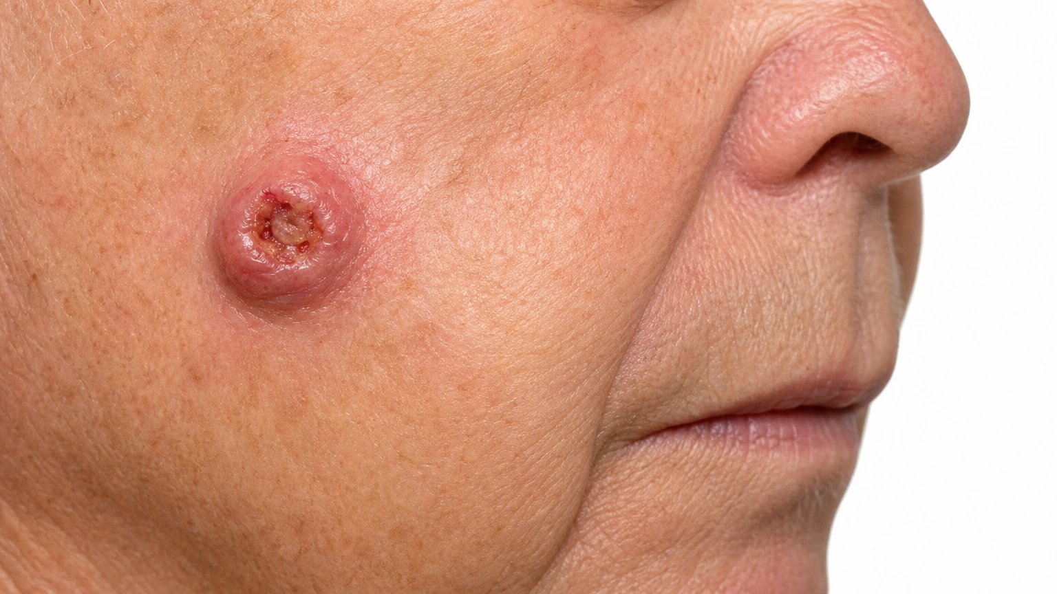
Dome-shaped red growth with a central keratin-filled crater on the cheek, representing keratoacanthoma.

Keratoacanthoma Fast-growing dome with a central crater.

Warts

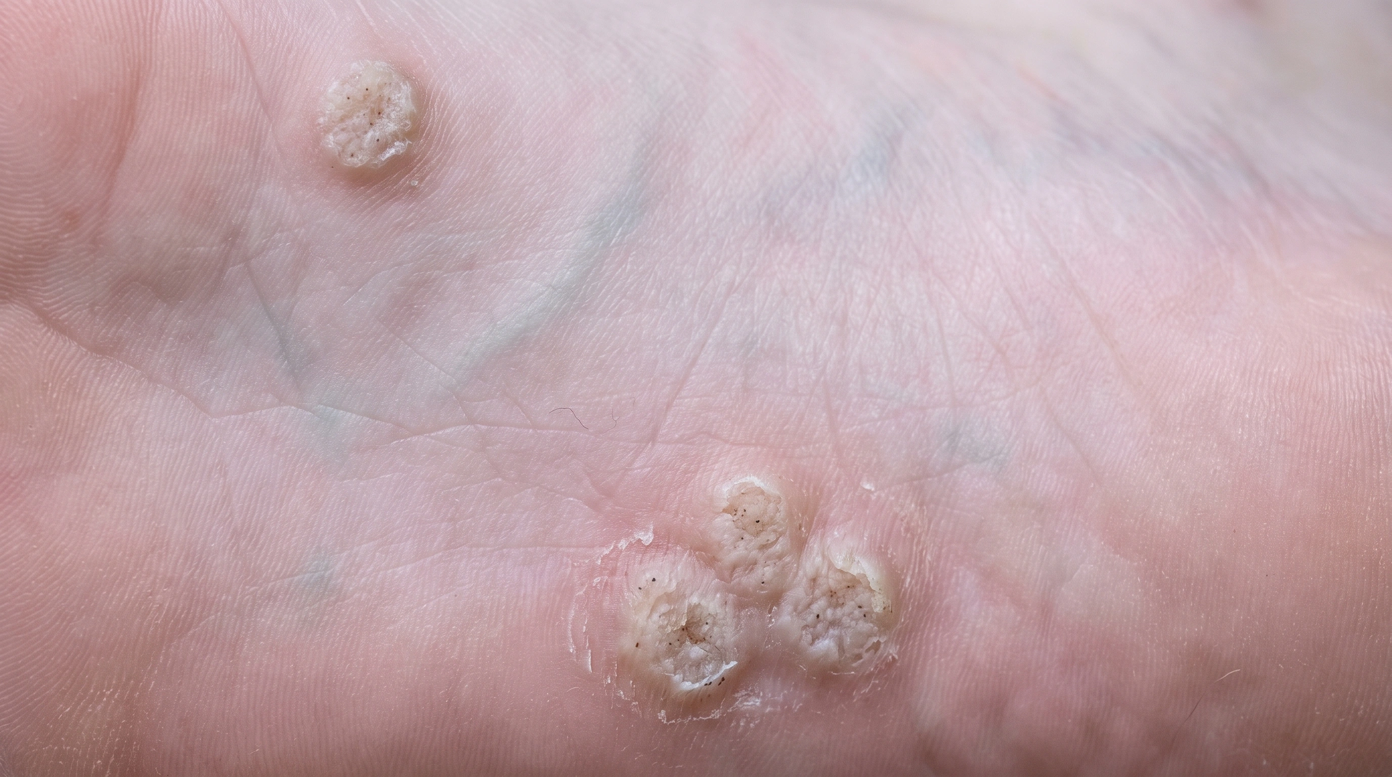
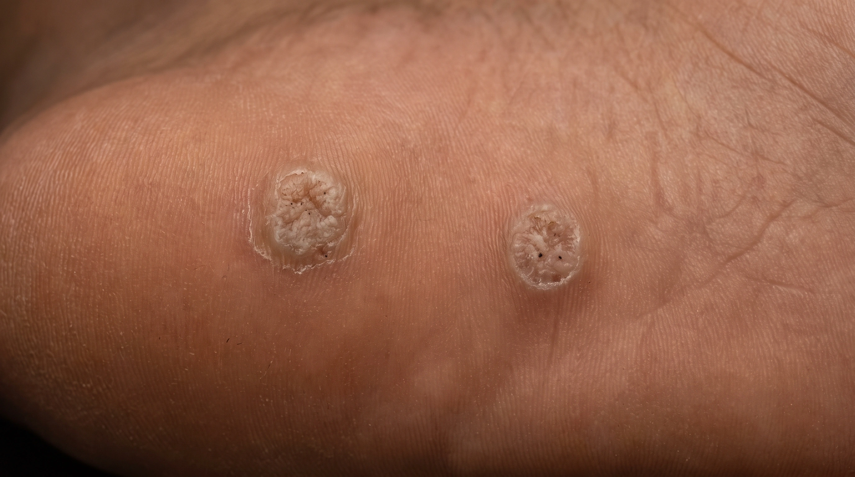
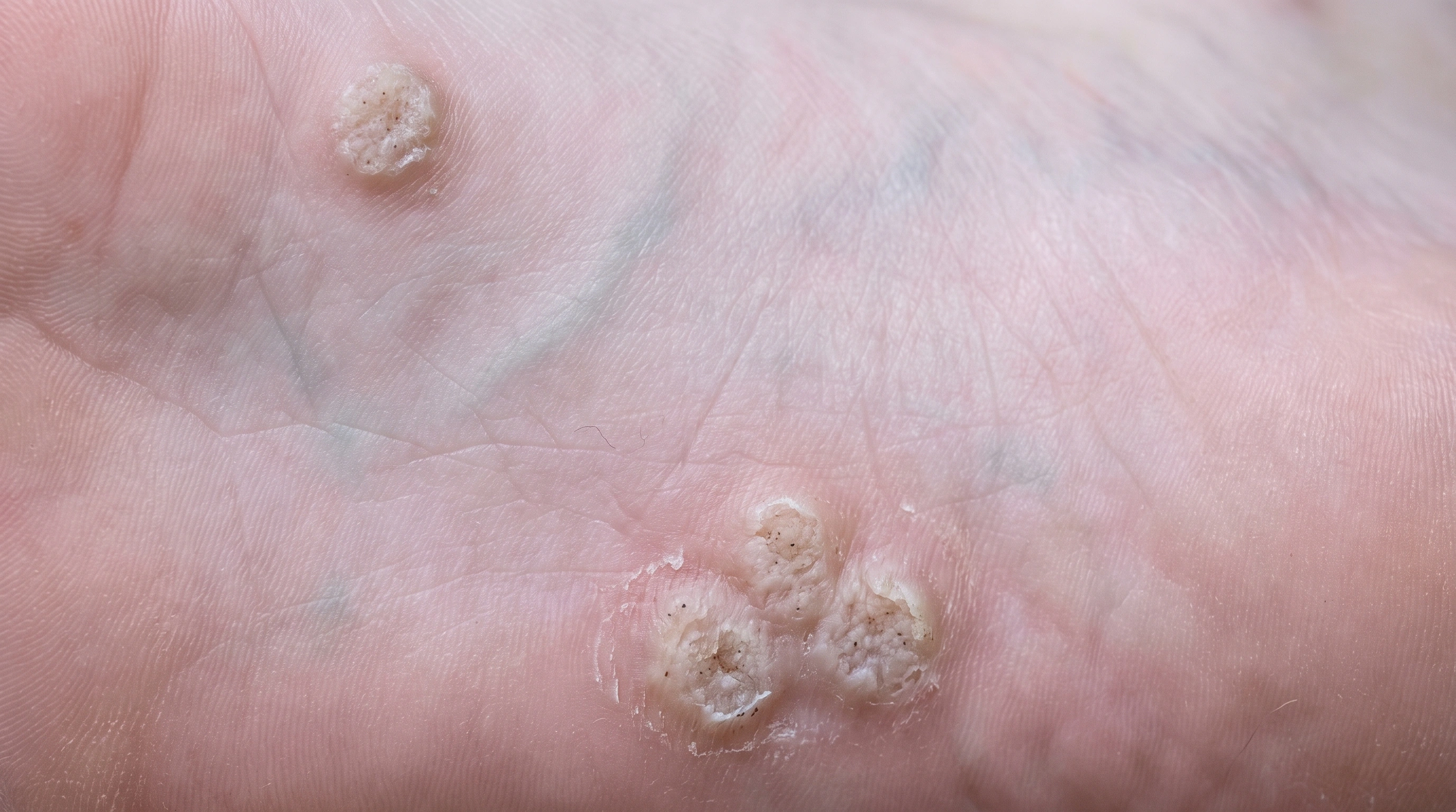
Warts: Black dots.

Common warts Rough bumps on hands and fingers.

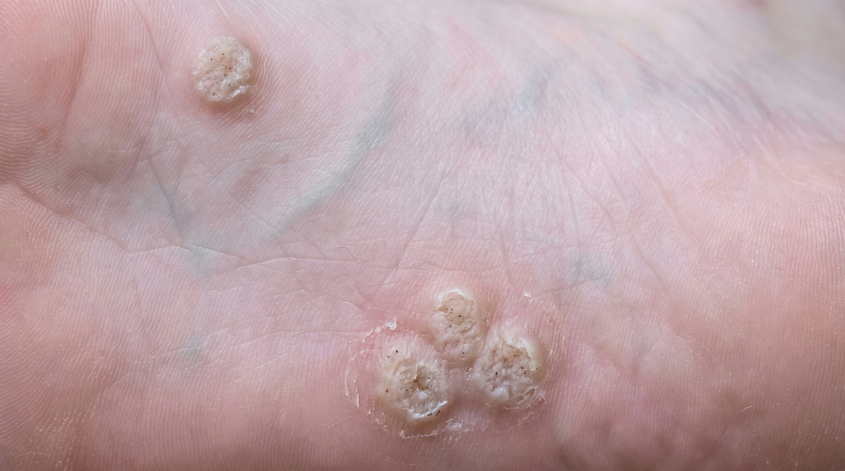
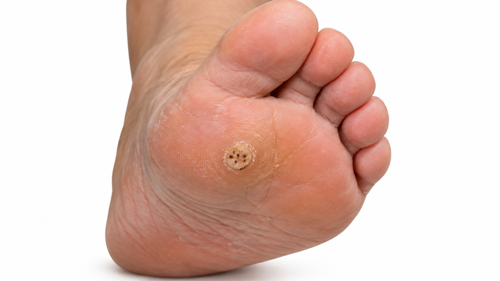
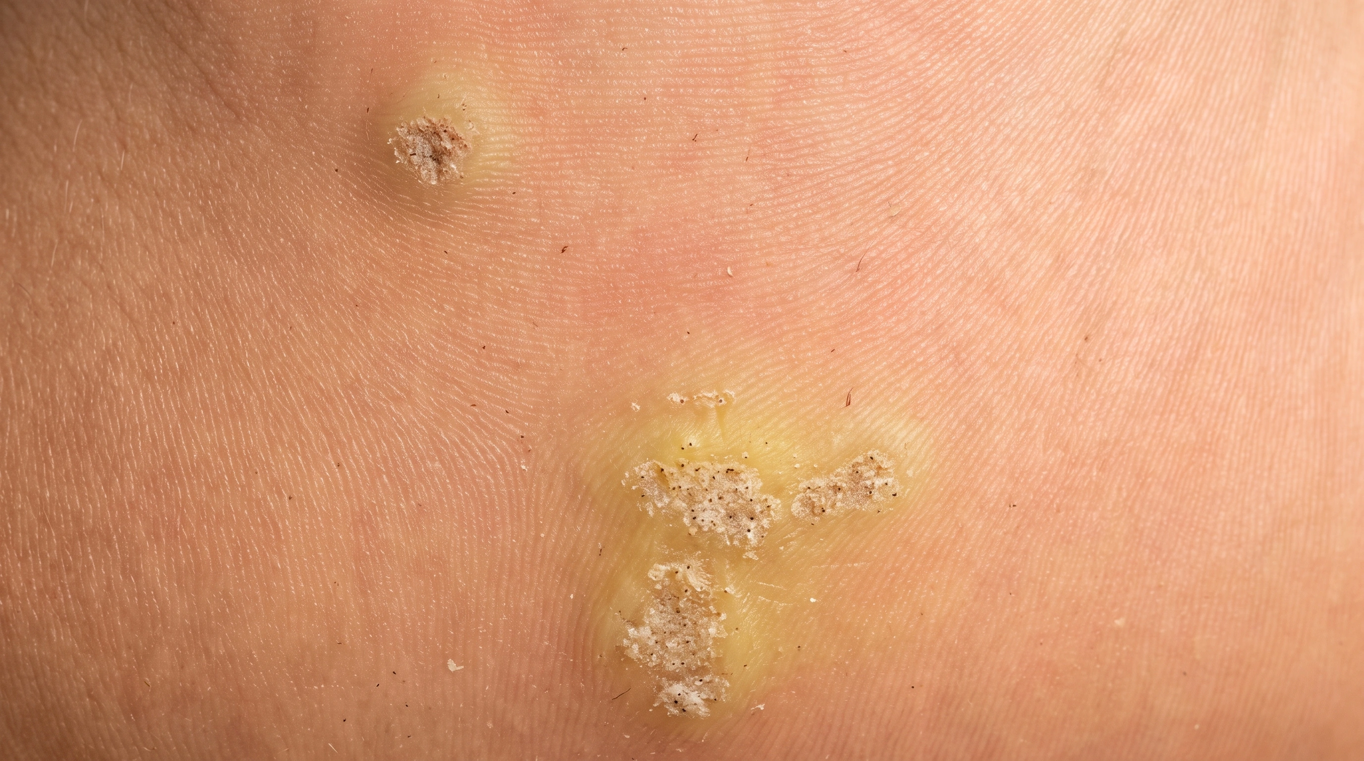
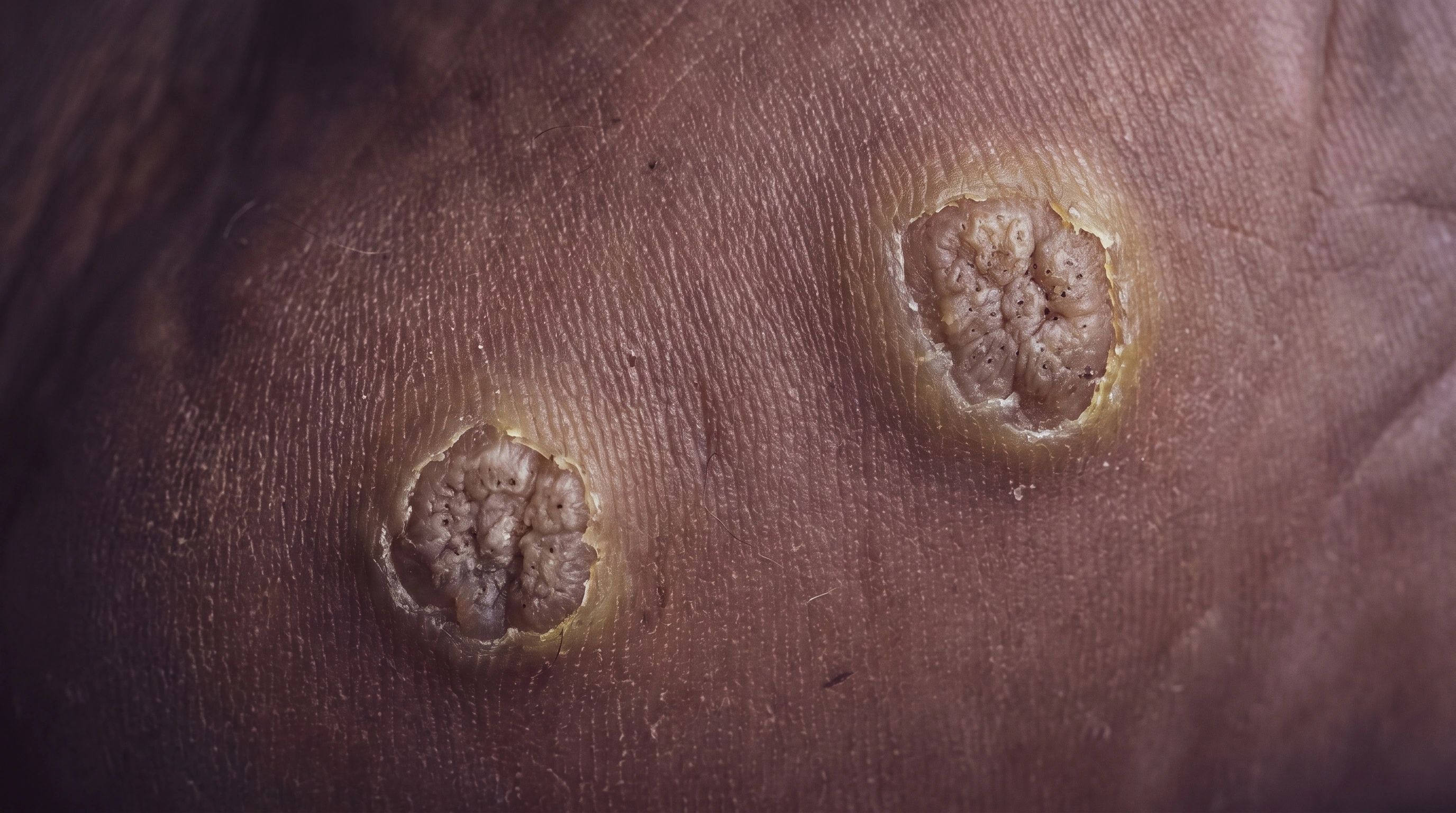
Warts: Plantar.

Plantar warts Flat, painful warts on the soles.

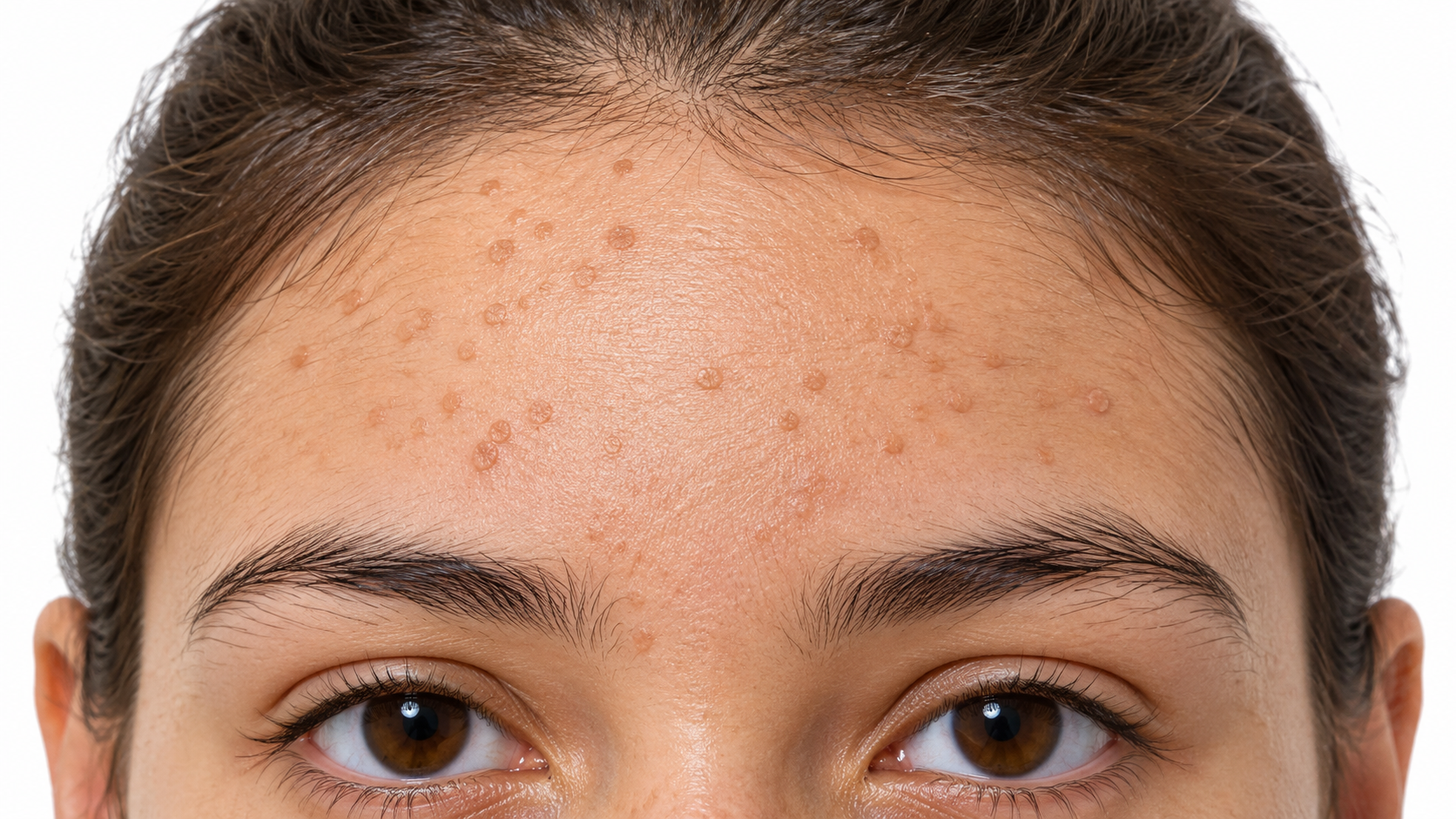
Warts: Flat.

Flat warts Small, smooth, flat-topped bumps.

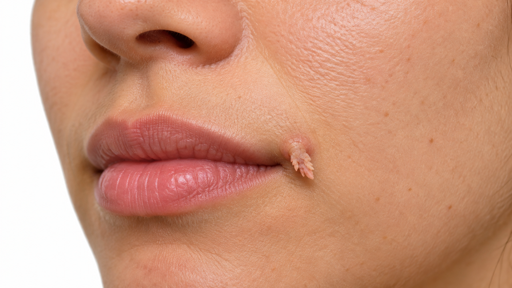
Warts: Filiform.

Filiform warts Long, thread-like growths on the face.

Skin tones
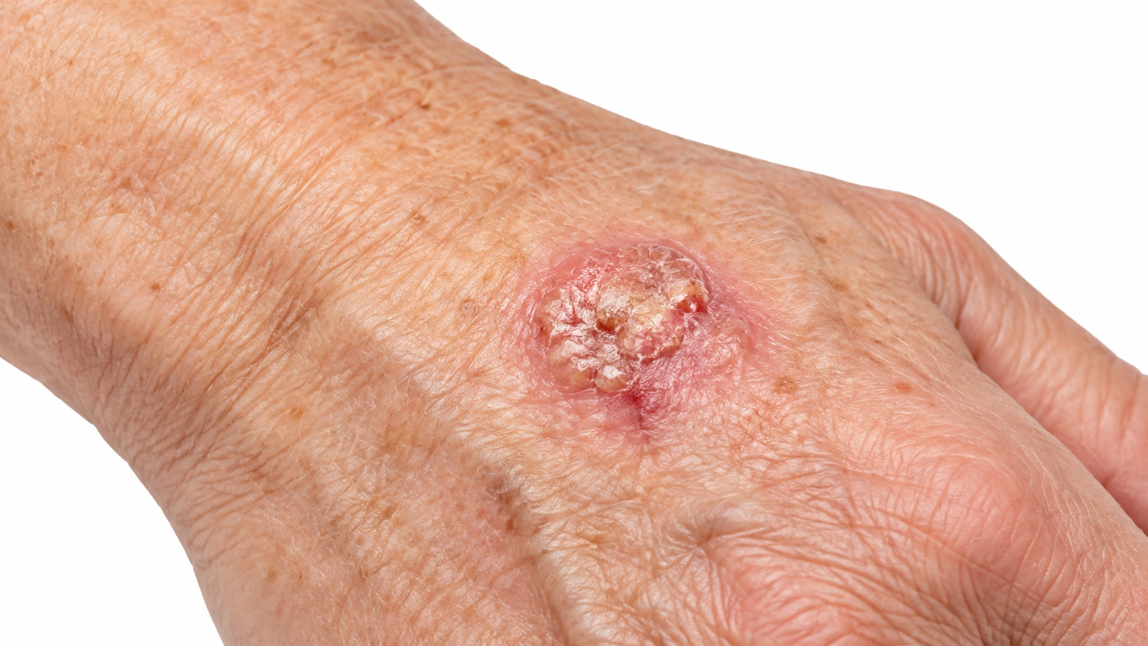
Crusted red raised lesion on a light-skinned sun-exposed hand.
Crusted sore on medium skin, representing squamous cell carcinoma.
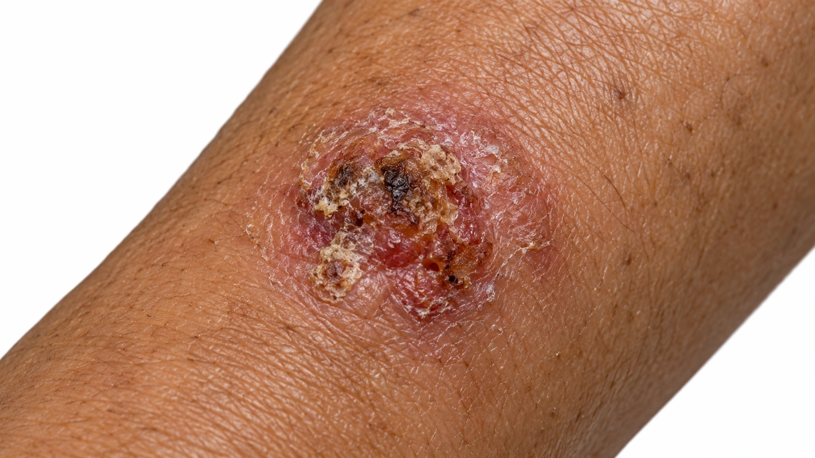
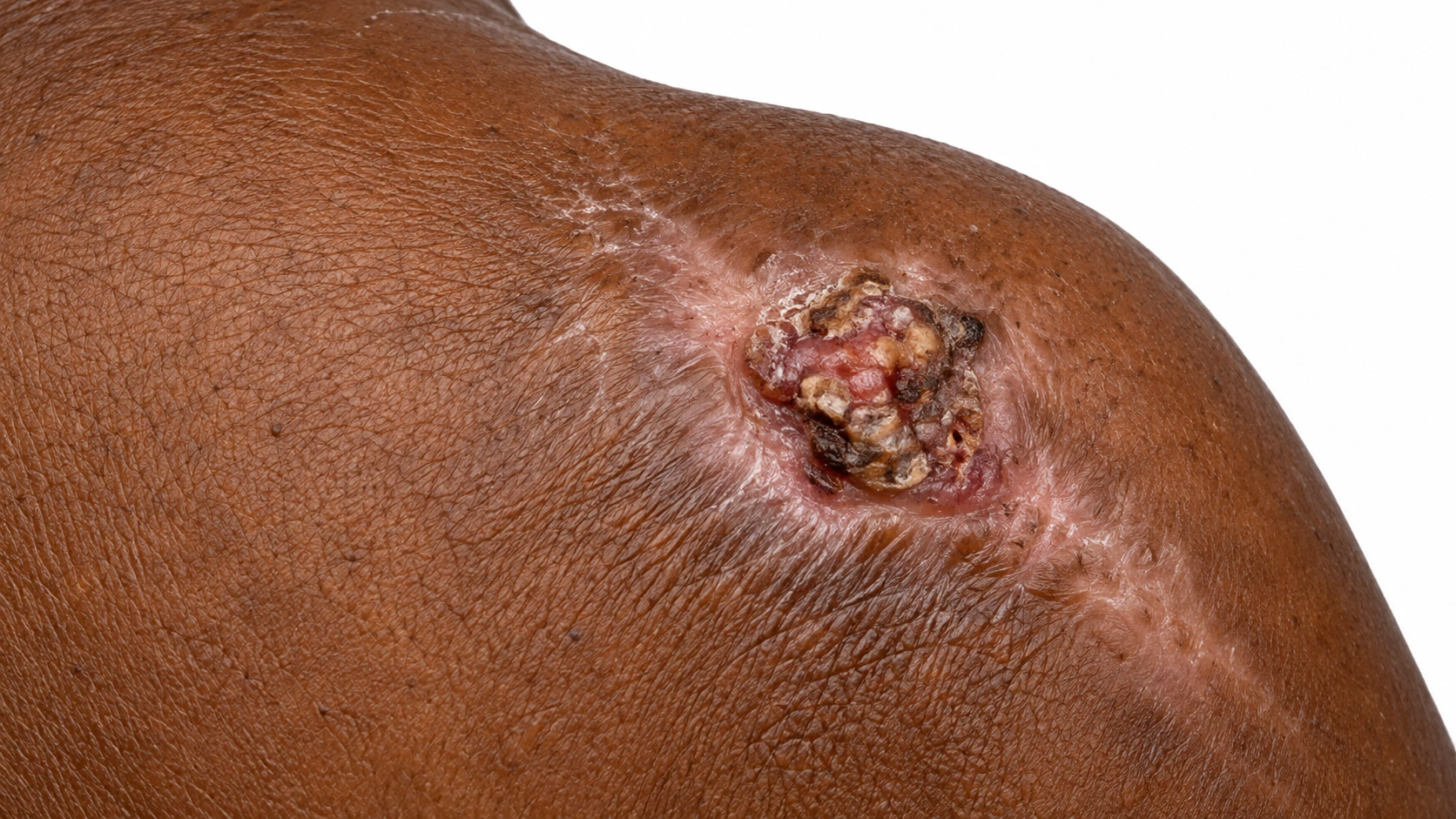
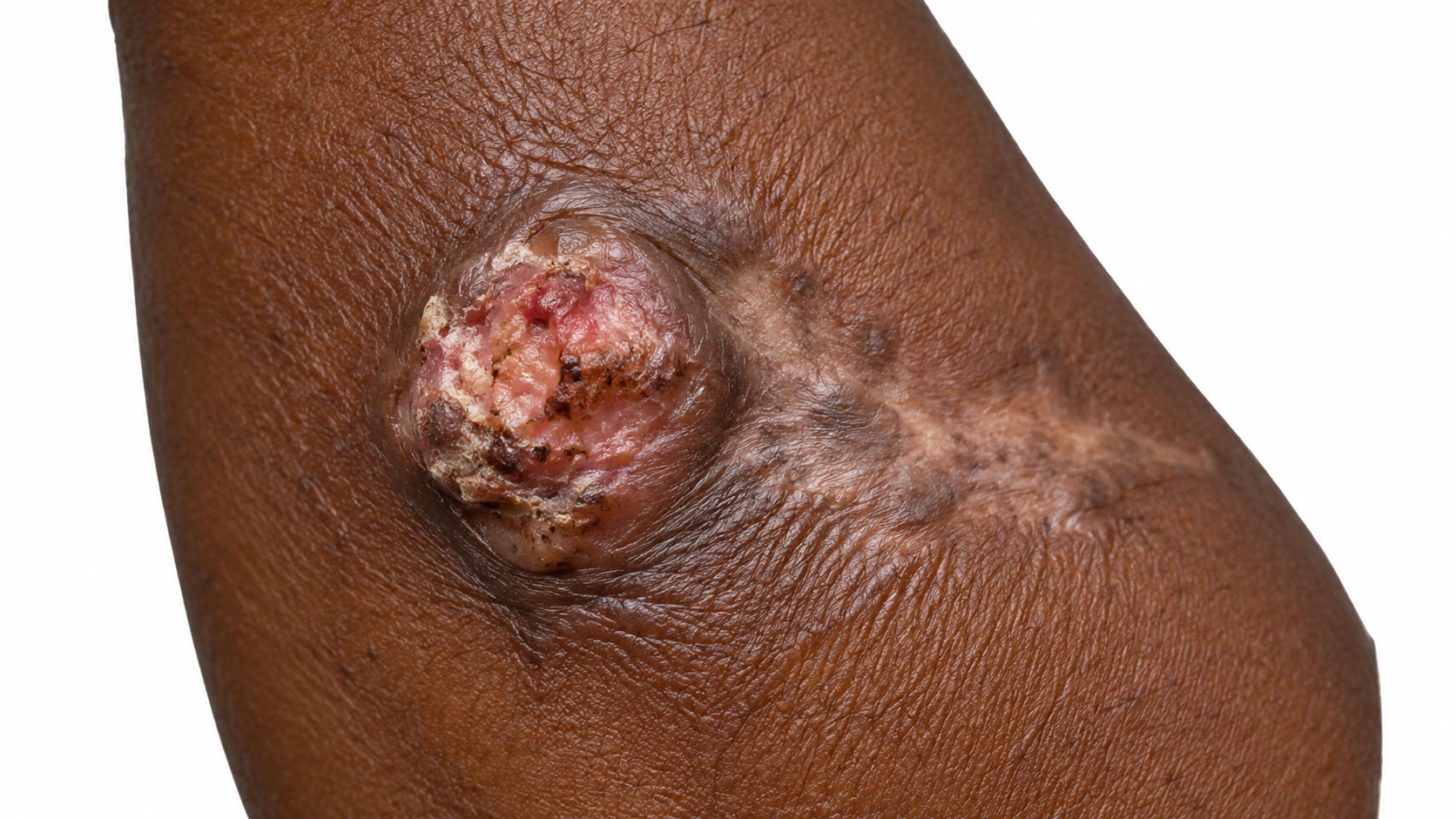
a crusted ulcerated lesion in scarred brown skin, representing squamous cell carcinoma.
a crusted raised lesion in scarred deep brown skin, representing squamous cell carcinoma.
A rough, dome-shaped wart on the side of a finger on fair skin. The bump is pinkish with a thin red ring at its base, and several tiny black dots are clearly visible in the thickened surface.
A rough, thickened wart on the back of a hand on medium olive skin. It is much the same color as the skin around it, with only a faint pink rim, and the fine skin lines stop at its border.
A firm, rough, gray-brown wart on the side of a finger on brown skin. There is no visible red ring around it, and the fine lines of the skin stop at the border of the bump.
Two rough, thickened warts on the back of a hand on deep brown skin. They are the same brown as the skin around them with no red rim, and they are visible mainly by their raised, uneven surface.
Diagram
Squamous Cell Carcinoma mechanism diagram. A rough or ulcerated growth forms: The surface may crust, bleed, or fail to heal. DNA damage changes skin cells: Ultraviolet exposure is a common cause. Cancer crosses into the dermis: Invasive cells grow below the epidermis.
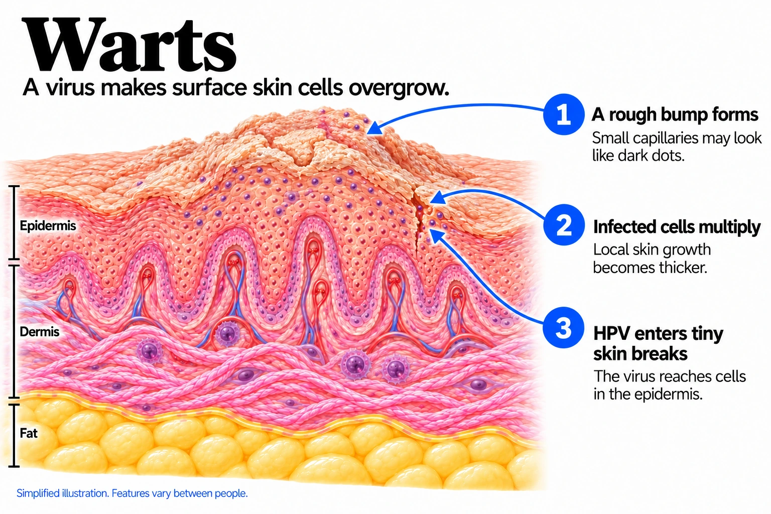
Warts mechanism diagram. HPV enters tiny skin breaks: The virus reaches cells in the epidermis. Infected cells multiply: Local skin growth becomes thicker. A rough bump forms: Small capillaries may look like dark dots.
In the skin

DNA changes allow abnormal surface skin cells to multiply, commonly after long-term ultraviolet damage. In invasive squamous cell carcinoma, the cancer grows through the epidermal boundary into the dermis.

Human papillomavirus enters through small skin breaks and stimulates local surface-cell growth. The thickened tissue forms a rough bump that may contain visible dark capillary dots.

Where it shows up

Often on sun-exposed skin
Hands and feet

When it usually starts

010203040506070+

Squamous Cell Carcinoma. Usually appears in older adults after cumulative sun damage; immunosuppression can lead to earlier disease.

Warts. Common in children and teenagers; adults can also develop them.

Causes and triggers

Causes

Squamous cell carcinoma starts when DNA damage makes skin cells grow out of control. Ultraviolet light is the main cause.

Warts are caused by the human papillomavirus infecting the top layer of skin. The virus makes skin cells grow faster, forming a rough bump.

Family tendency

Fair skin that burns easily is inherited and raises the risk.

Risk factors
Fair skin, Weakened immunity, Actinic keratoses, Long-standing wounds or scars
Children and teens, Weakened immunity, Nail biting
How it spreads
  • Skin-to-skin contact — Well established
  • Cuts, scrapes and hangnails — Strong link
  • Picking or biting warts — Strong link
  • Shared towels or razors — Possible
Trigger
● Squamous Cell Carcinoma
● Warts
Unprotected sun exposure
Well established
Not listed
Tanning beds
Well established
Not listed
HPV in some areas
Strong link
Not listed

Daily Care

Squamous Cell Carcinoma
Warts
Daily care

Habits that help

  • Daily broad-spectrum sunscreen
    SPF 30 or higher on face, ears, neck and hands.
  • Clothing, hats and shade
    More than sunscreen alone, and no reapplication.
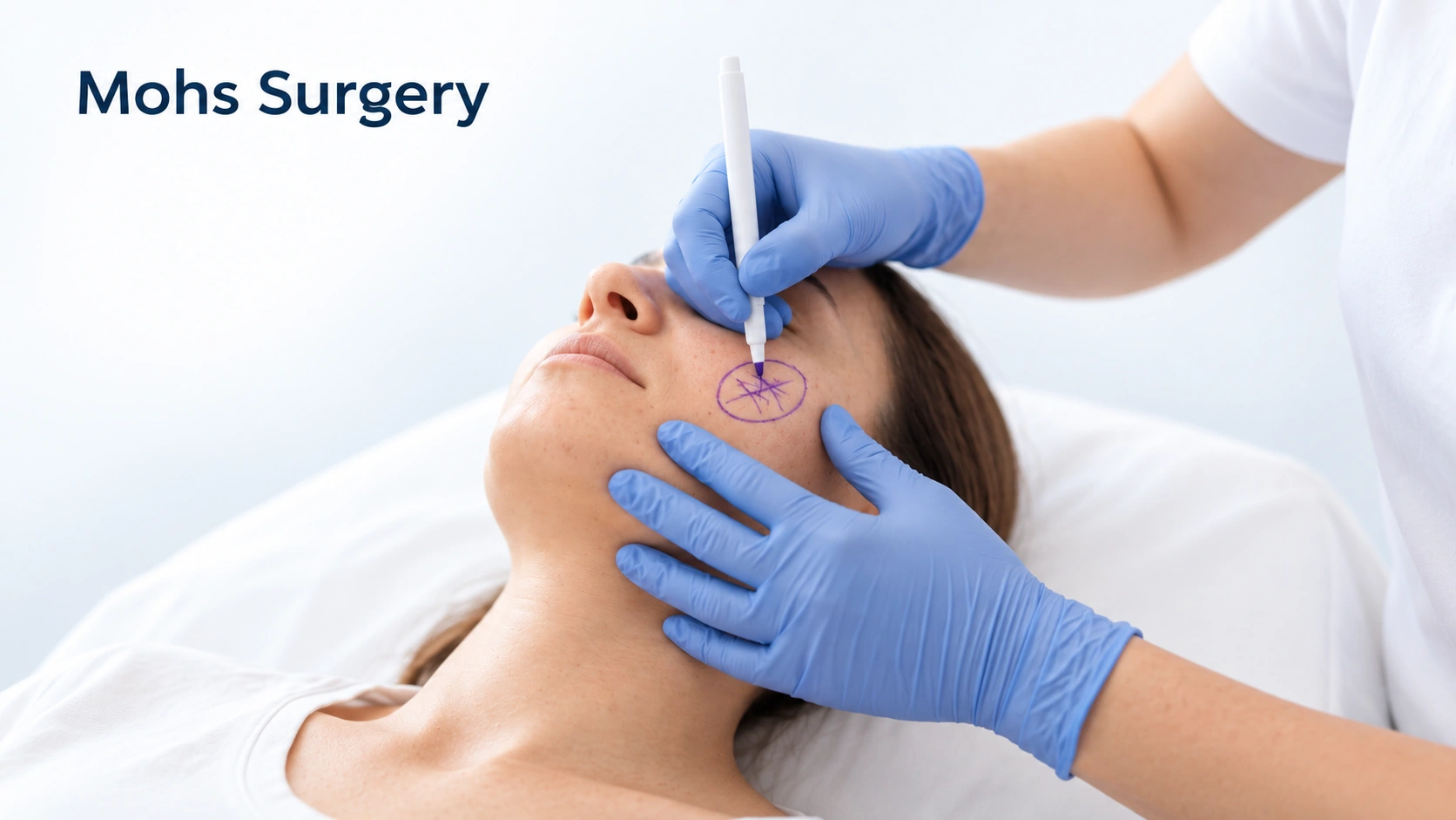
  • Mohs surgery
    Layers checked during the appointment - spares tissue, highest cure rate.
  • Curettage and electrodesiccation
    Small, low-risk tumors, not the face.
  • Radiation when surgery is not possible
    Also used after aggressive tumors.
  • Prescription creams for the earliest forms
    Fluorouracil and imiquimod treat in situ disease and actinic keratoses, not invasive tumors. If you are pregnant or breastfeeding, ask the doctor managing your pregnancy.
  • Treating actinic keratoses
    Fewer of these cancers later.
  • Nicotinamide
    500 mg twice a day modestly reduces new skin cancers.
  • Regular skin checks after treatment
    How the next one is caught small.
  • Discuss closer monitoring if your immune system is suppressed
    Faster, more aggressive tumors - ask your transplant team about surveillance, and never stop medication yourself.
  • Arrange prompt assessment for a tumor on the lip or ear
    Higher risk of spread.

Avoid

  • Avoid: Continuing unprotected sun exposure
    Each dose raises the next cancer's odds.
  • Avoid: Tanning beds
    A recognized cause, pointless after a diagnosis.
  • Avoid: Ignoring actinic keratoses
    The step immediately before this cancer.
  • Avoid: Black salve and escharotic pastes
    Destroy the surface, leave tumor at depth.
  • Avoid: Picking the crust off repeatedly
    Keeps it looking nearly healed.
  • Avoid: Smoking
    Lip cancer, and poorer healing after removal.
  • Avoid: Waiting on a spot that will not heal
    The biggest factor in outcome.
  • Avoid: Skipping follow-up appointments
    New cancers are common after the first.

Habits that help

  • Covering the wart
    Keeps virus off other spots and holds treatment against the skin.
  • Keeping hands and feet dry
    Change socks during the day, dry between the toes, air your shoes.
  • Your own kit
    Flip-flops in shared showers, own towel, own clippers.
  • Follow safe filing instructions for wart treatment; avoid injured or high-risk feet
    Use only gentle removal of dead surface skin when advised; never cut or file to pain or bleeding. High-risk feet need professional advice.
  • Cantharidin, applied in clinic
    A blistering agent, painless going on, so it suits children.
  • Immune-based treatments
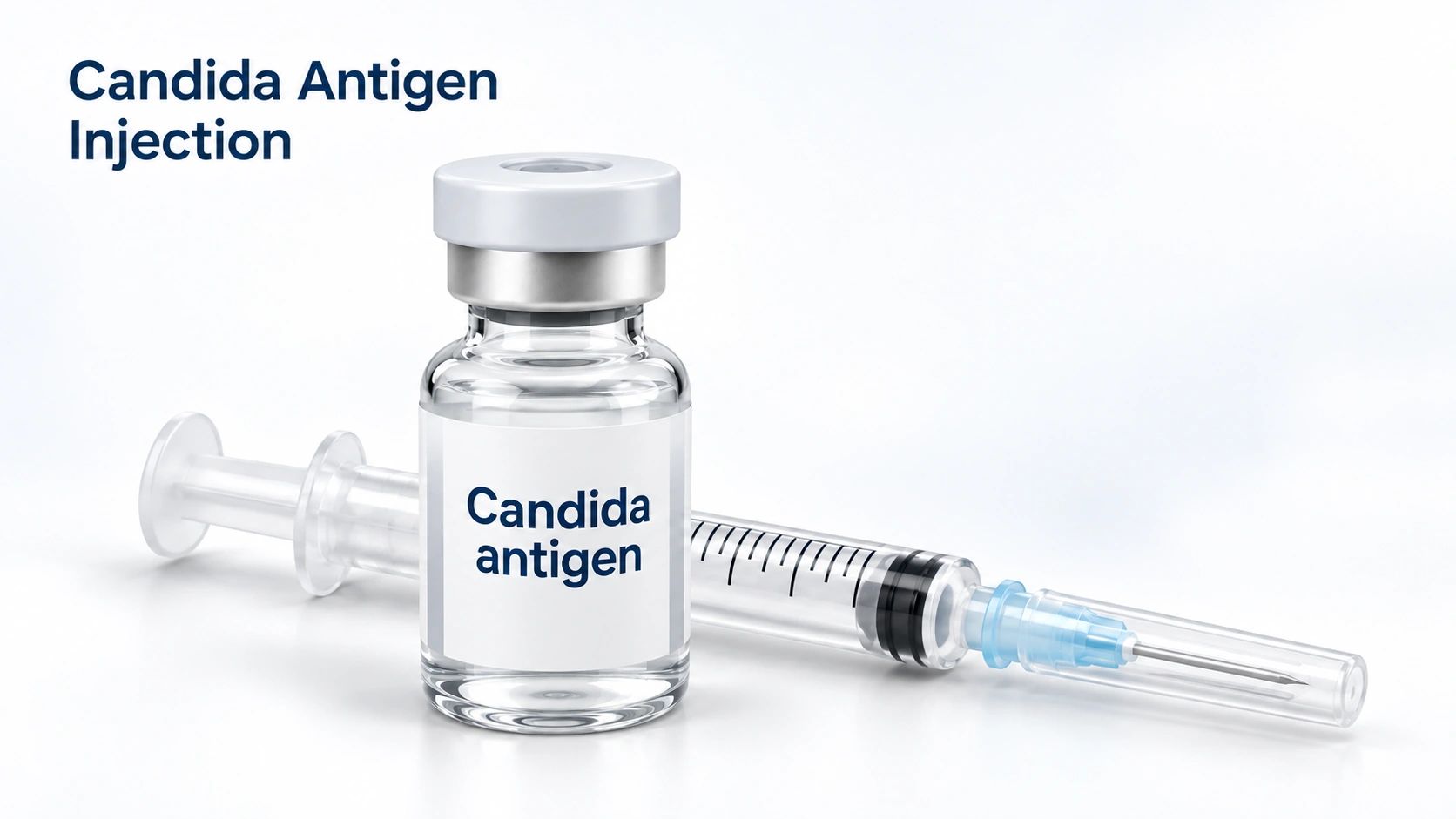
    Imiquimod cream or candida antigen injections for warts that keep returning.
  • Treating dry skin and eczema properly
    Intact, moisturized skin is much harder to enter.
  • Allow the treatment-specific course and review if it is not helping
    The required course varies by treatment and site. Review persistent, painful or uncertain lesions rather than repeatedly damaging the skin.
  • Review immune-suppressing medicines with the prescriber; do not stop them yourself
    Immune suppression can affect infection persistence. Tell the treating clinician and do not stop a prescribed medicine on your own.
  • Follow safe wart preparation advice; do not file to pain or bleeding
    Use only gentle removal of dead surface skin when the product recommends it. Never file to live, painful or bleeding skin. Do not share files; get advice for diabetes, poor circulation or reduced sensation.

Avoid

  • Avoid: Picking, biting and pulling at warts
    Carries virus to fingers and lips, and leaves broken skin for it.
  • Avoid: Shaving over a wart
    Seeds virus along the whole shaving path.
  • Avoid: Walking barefoot in shared showers
    Warm wet floors are where the virus survives longest.
  • Avoid: Skin that stays wet for hours
    The outer layer softens and stops being a seal.
  • Avoid: Nail biting and picking at hangnails
    The most reliable way to get warts around the nails.
  • Avoid: Sharing personal tools
    Towels, razors, nail clippers, pumice stones and files move the virus.
  • Avoid: Reusing one file on several warts
    It spreads them between your own sites.
  • Avoid: Stopping treatment too early
    Virus stays at the edges a week or two after the surface looks normal.
  • Avoid: Eczema, dry skin and cracked heels
    Broken skin is an open door.

Treatment differences

Evidence labels come from each condition’s own treatment list.

Treatment
● Squamous Cell Carcinoma
● Warts
Filter by type
Diagnosed squamous cell carcinoma type

Select one or more.

Wart type

Select one or more.

Over-the-counter
None listed
Existing illustration of Home cautery pens and freeze-off kits.

Home freeze kits

Limited evidence

The pharmacy sprays get nowhere near as cold as clinic liquid nitrogen, so they work best on small, new, thin warts on the hands and poorly on thick foot warts. They sting and can blister. On deeper skin tones they carry the same risk of leaving a pale or dark mark as clinic freezing does.

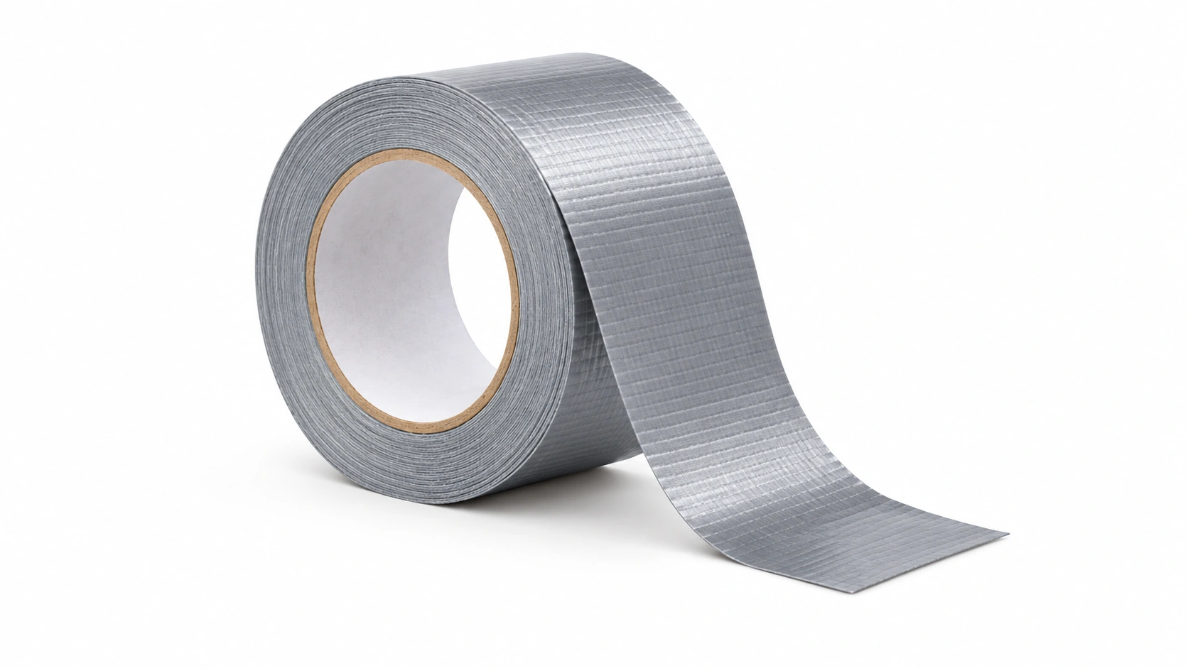
Existing Dermapedia illustration of duct tape.

Duct tape

Limited evidence

Covering a wart with tape for a few days at a time, then filing it, is cheap and harmless, and the trials are genuinely mixed on whether the tape itself adds anything. Any benefit is probably from the covering and the filing. It is reasonable alongside salicylic acid and a poor substitute for it.

Paula's Choice 2% BHA exfoliant, La Roche-Posay Effaclar cleanser and a CeraVe psoriasis cream pump

Salicylic Acid

Strong evidence

The first treatment for almost every ordinary hand or foot wart, and the one with the best evidence outside the clinic. It dissolves the infected thickened skin a layer at a time, so it only works if it goes on every single day. Soak the wart for five minutes, file the dead surface off, apply, and cover it. Twelve weeks is a fair trial; ten days is not.

Prescription topicals
None listed
Illustration: Generic topical cream tube and white cream.

Imiquimod

Limited evidence

Imiquimod cream. A cream that works on the last step of the problem: it makes the local immune system pay attention to the virus. It is used mainly for flat warts on the face and for warts that keep coming back, applied a few nights a week for weeks. It reddens and irritates the skin while it works, and it does poorly on thick foot warts.

Tretinoin — Header Image

Tretinoin

Limited evidence

A retinoid cream used for the flat warts that appear in groups of dozens on the face and the backs of the hands, where acid and freezing would be too harsh over so wide an area. It speeds up the turnover of the infected surface cells. It takes months, and it makes the skin dry and sun-sensitive while it runs.

Fluorouracil

Limited evidence

5-fluorouracil cream. A chemotherapy cream at a low strength, applied to the wart itself, often under tape. It is used for flat warts and for warts that have not shifted with acid, and it is a common choice in children because it does not hurt going on. It reddens and can crust the treated skin, and it is not used in pregnancy.

Prescription pills
None listed
An illustrative bottle labeled Cimetidine beside plain tablets.

Cimetidine

Limited evidence

An old stomach-acid tablet that people ask about for multiple stubborn warts, on the idea that it nudges the immune system. The better-controlled trials found it no more effective than a dummy tablet in adults. It is safe and cheap, so it is occasionally still tried in children, but it should not replace treatment that is known to work.

Injections
None listed
Editorial illustration for Candida Antigen Injection, based on the entry’s treatment or equipment reference.

Candida Antigen Injection

Limited evidence

Candida antigen injection. A tiny amount of yeast protein injected into one wart, to wake the immune system up in that spot. What makes it useful here is that untreated warts elsewhere on the body often clear at the same time, so it suits someone with many warts rather than one. It is repeated every few weeks, and it can cause a day or two of aching or flu-like feeling.

Bleomycin Injection

Bleomycin Injection

Limited evidence

Bleomycin injection. Injected directly into warts that have resisted everything else, particularly thick ones on the soles and around the nails. Clearance rates are good. It is painful enough that the area is numbed first, it can leave a permanent nail change if used at the nail fold, and it is not used in pregnancy, so it sits late in the order for a reason.

Procedures
Editorial illustration for Mohs Surgery, based on the entry’s treatment or equipment reference.

Mohs Surgery

Strong evidence

The tumor is removed a layer at a time and each layer is checked under the microscope during the same appointment, so the surgeon stops as soon as the edges are clear. It gives the highest cure rate and takes the least normal skin, which is why it is used on the face, ears, lips, hands, feet and genitals, and for large, recurrent or aggressive tumors. It is a long appointment rather than a difficult one.

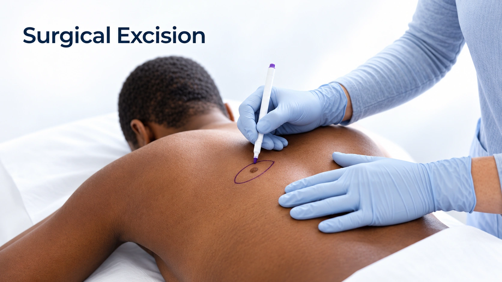
Editorial illustration for Surgical Excision, based on the entry’s treatment or equipment reference.

Surgical Excision

Strong evidence

The standard removal: the tumor is cut out with a margin of normal skin around it and the wound is stitched closed, usually in one visit under local anesthetic. It cures the large majority of straightforward tumors on the trunk, arms and legs. The margins are checked afterwards rather than during, so occasionally a second procedure is needed to clear an edge.

Editorial illustration for Electrodesiccation and Curettage, based on the entry’s treatment or equipment reference.

Electrodesiccation and Curettage

Moderate evidence

The tumor is scraped out with a small loop and the base is sealed with an electric current, repeated a few times in one sitting. It is quick and effective for small, thin, low-risk tumors on the trunk and limbs. It is not used on the face, on large or recurrent tumors, or anywhere depth matters, because nothing is sent for margin checking and it leaves a round pale scar.

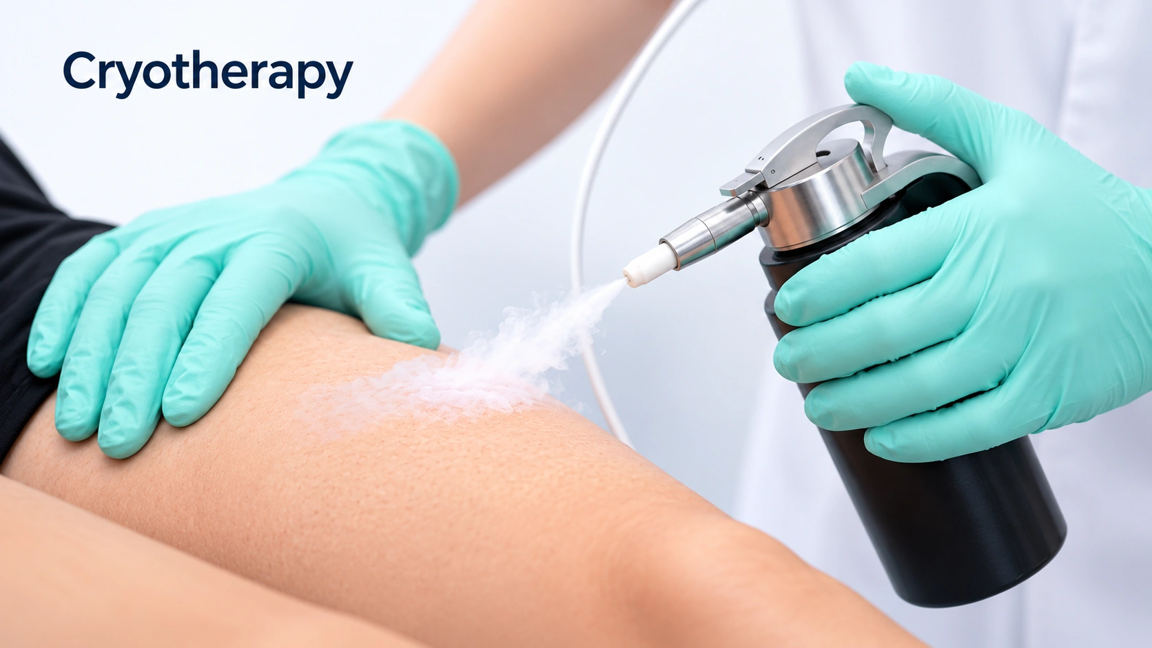
Editorial illustration for Cryotherapy, based on the entry’s treatment or equipment reference.

Cryotherapy

Moderate evidence

A quick freeze in clinic, used for actinic keratoses and occasionally for a small patch of carcinoma in situ. It is a treatment for the step before this cancer, not for the cancer itself. Freezing an invasive tumor leaves cells behind at depth and hides what is happening under a healing crust.

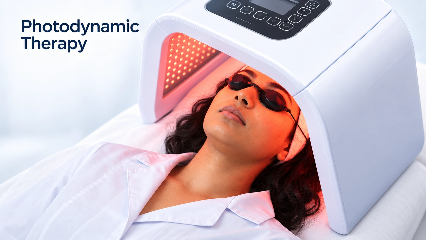
Editorial illustration for Photodynamic Therapy, based on the entry’s treatment or equipment reference.

Photodynamic Therapy

Limited evidence

A light-activated cream treatment for the earliest forms only — actinic keratoses and some cases of carcinoma in situ. It treats a whole patch of sun-damaged skin at once and heals with a good cosmetic result. It has no role in an invasive tumor, and using it on one delays proper removal.

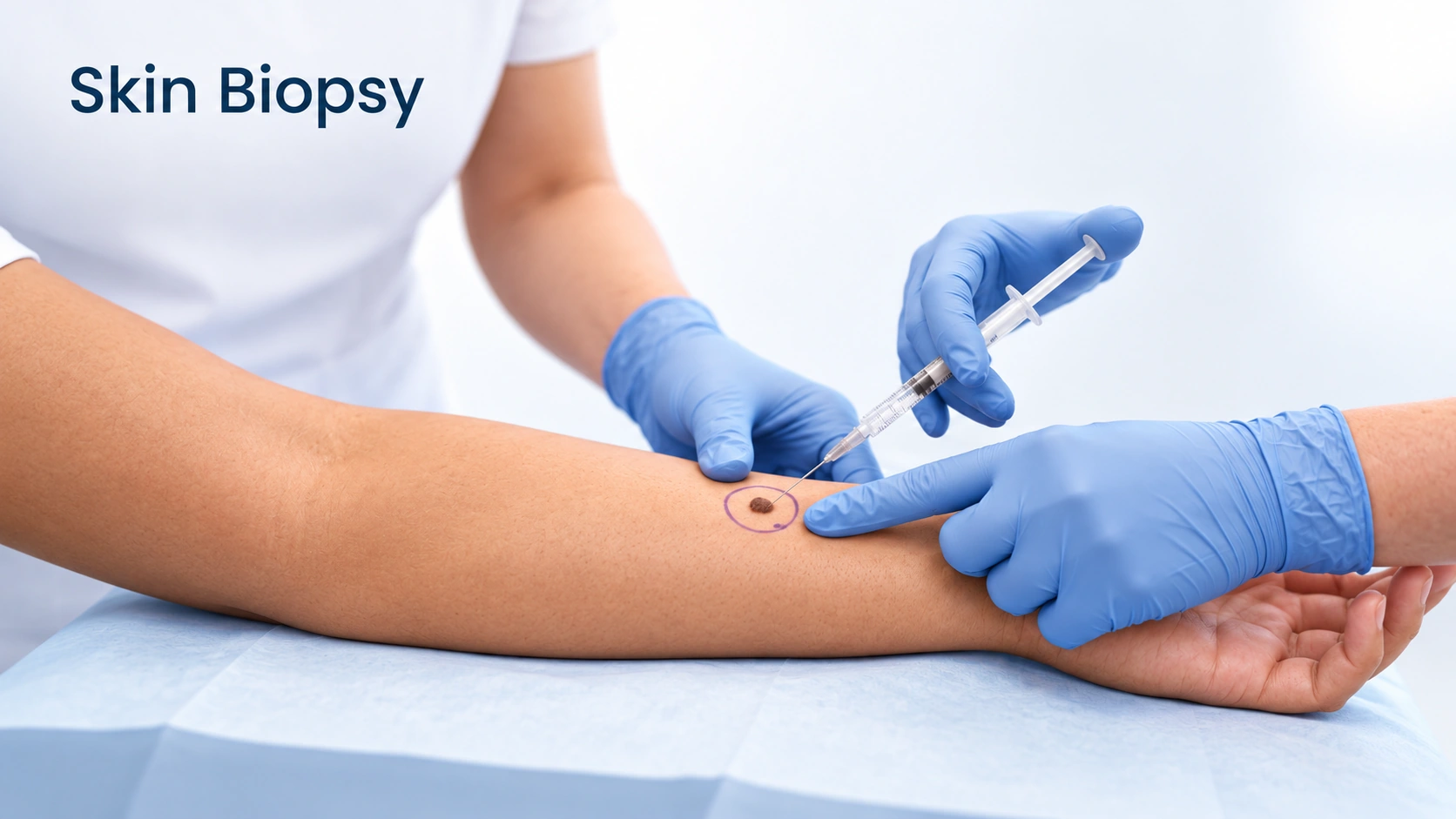
Editorial illustration for Skin Biopsy, based on the entry’s treatment or equipment reference.

Skin Biopsy

Listed

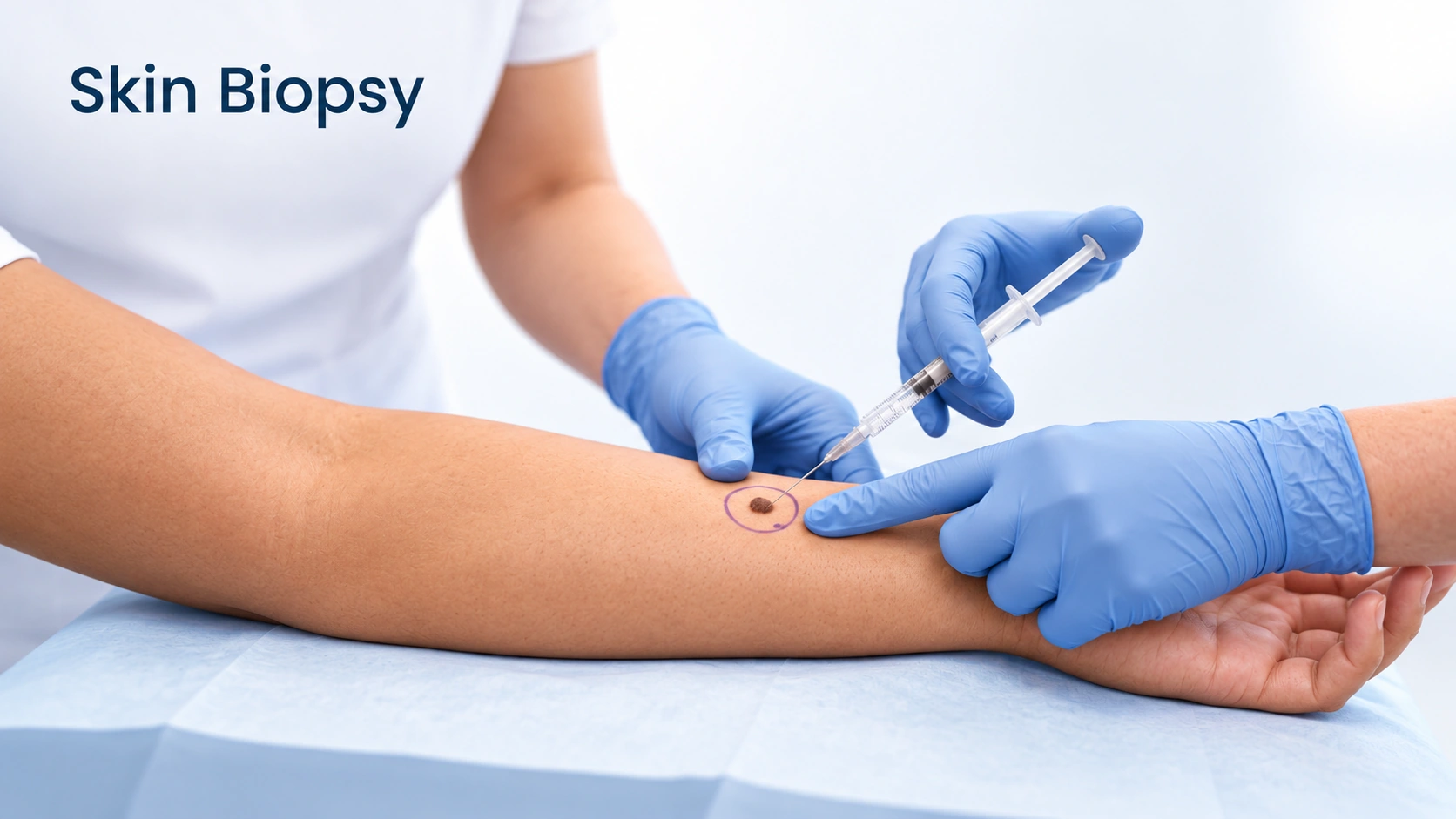
Nothing else on this page happens without it. Even an experienced dermatologist with a dermatoscope cannot settle this by looking, and the report says far more than yes or no — how deep it goes, how aggressive the cells look, whether it is tracking along a nerve. Those details choose the treatment. It takes a few minutes under local anesthetic and leaves a small wound. A biopsy does not make cancer spread.

Editorial illustration for Electrodesiccation and Curettage, based on the entry’s treatment or equipment reference.

Electrodesiccation and Curettage

Limited evidence

The wart is numbed, burned with an electric current and scraped away in one visit. It is kept for a single stubborn wart that has failed everything gentler, because it leaves a scar, and a scar on the sole of a foot can hurt more than the wart did. Warts can still regrow at the edge of the scar.

Editorial illustration for Cryotherapy, based on the entry’s treatment or equipment reference.

Cryotherapy

Moderate evidence

Liquid nitrogen freezes the infected skin and the injury also draws immune attention to the spot. It needs repeating every two to four weeks, usually three or four times, and a wart regrowing from the edge between sessions is normal rather than failure. Over several months its results are broadly similar to salicylic acid used properly. It stings, it blisters, and on deeper skin tones it can leave a lasting pale or dark mark.

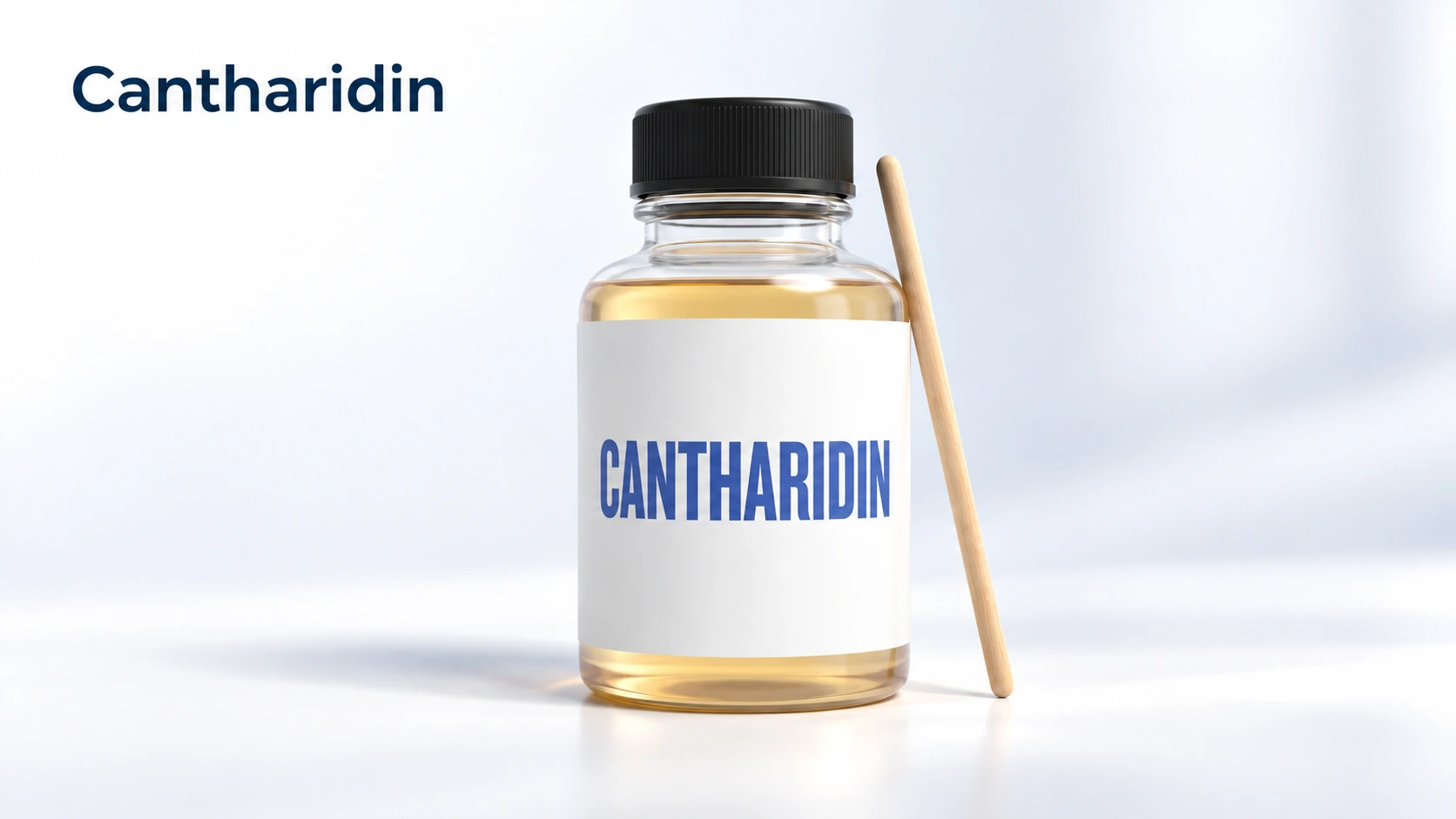
Editorial illustration for Cantharidin, based on the entry’s treatment or equipment reference.

Cantharidin

Limited evidence

A blistering liquid painted on in clinic and washed off a few hours later. It is painless going on, which is why it is a common first choice for children and for warts on the face. The blister that follows lifts the infected skin away over the next few days and can be sore. It usually takes more than one visit.

Paring

Limited evidence

Paring in clinic. The top dead layer is shaved off with a blade. It does not hurt, because that layer has no nerve supply, and it does two jobs at once: it confirms the diagnosis by exposing the tiny clotted vessels a callus does not have, and it lets acid or liquid nitrogen reach live infected skin. It is done alongside another treatment, never as the whole treatment.

V-Beam

Limited evidence

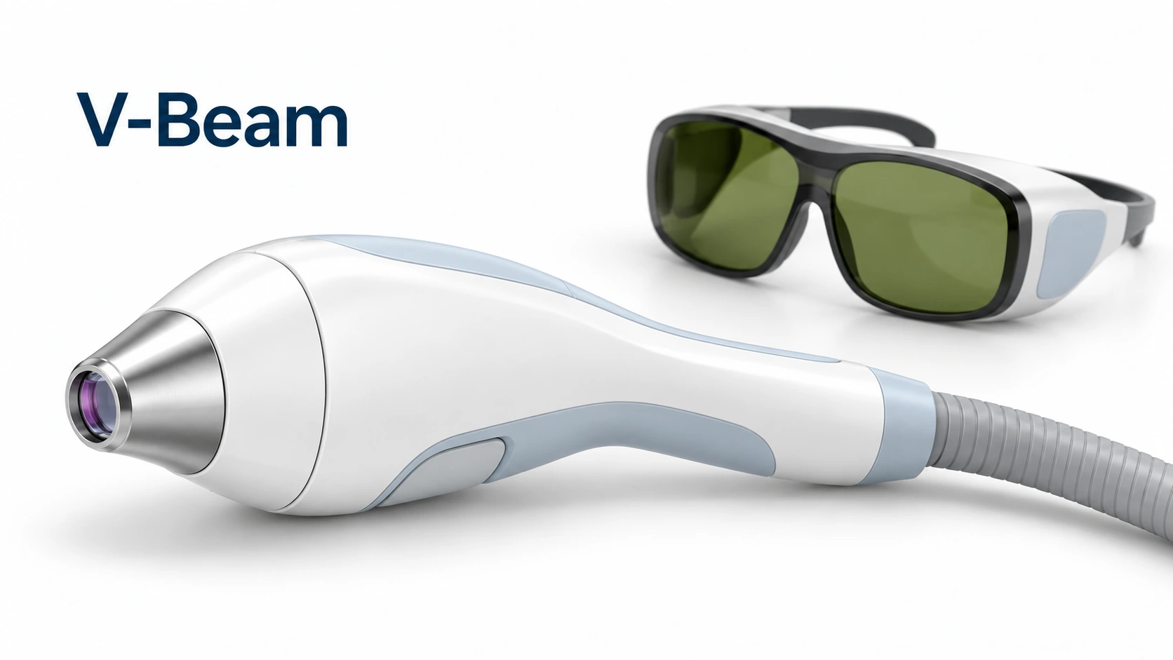
The laser targets the small blood vessels feeding the wart rather than the virus itself. It is an option for stubborn warts on the hands and around the nails when freezing has not worked, and it spares the skin around it, which matters on the face. Several sessions are needed, results are inconsistent, and it is not usually covered by insurance.

Outlook

Long term

Most skin squamous cell carcinomas are cured with appropriate treatment, but they should not be left untreated. Some grow or spread more quickly, especially with immune suppression. Follow-up helps detect recurrence and additional skin cancers.

Warts are self-limiting — they end on their own eventually. In children, roughly two thirds clear within two years without treatment. Adults are slower, and warts present for several years are least likely to go quietly.

Complications

Possible complications
  • A small percentage reach the lymph nodes
More details

A small percentage reach the lymph nodes
A small percentage spread to nearby lymph nodes and occasionally further. Higher risk with large or deep tumors, ear or lip sites, recurrence, and immunosuppression.

  • Spreading to new spots
  • Color change after treatment
  • Warts around and under the nails
More details

Spreading to new spots
An untreated wart seeds others nearby, so one bump becomes a ring of small ones around it, or a line of them along a scratch or a shaving path. Picking, biting and filing several warts with the same file are the usual ways it happens. Covering the wart, keeping your own file and treating early keep the number down.

Color change after treatment
Freezing, and any treatment that blisters or burns, works by injuring the skin, and injured skin can heal a different color. A pale ring around the treated spot can last a year or more and is sometimes permanent, and a dark mark can outlast the wart by months. Both are far more likely on deeper skin tones, which is why gentler treatment and a small test spot are often the better trade.

Warts around and under the nails
Warts that set up in the nail folds are the most stubborn of the group, because the nail shields the virus from creams and from freezing. Left long enough, a wart growing under the nail can lift it or leave a permanent ridge or split in the nail plate. Nail biting and picking at hangnails are how most of them start, and they are the ones most worth taking to a dermatologist early.

When to get help

When to get help

Arrange prompt assessment of a persistent scaly, crusted, enlarging or non-healing lesion, especially if tender or bleeding.

Seek assessment for an uncertain, painful, bleeding or changing growth, or persistent warts with weakened immunity. Genital lesions need separate assessment.

Common questions

Squamous Cell Carcinoma:Is squamous cell carcinoma serious?

It is a real cancer and treated as one. Most cases are cured by removal, which is why the emphasis is on early checks. Untreated, it grows into deeper structures and a small share reach lymph nodes. How serious it gets depends on how long it is left.

Squamous Cell Carcinoma:How fast does it grow?

Usually weeks to months rather than days or years. The keratoacanthoma variant can appear and enlarge within a few weeks. In immunosuppressed people growth can be much faster, which is why the advice for that group is more urgent.

Squamous Cell Carcinoma:Will I need Mohs surgery?

It depends on the site and how the tumor looks under the microscope. Mohs is generally used on the face, ears, lips, hands, feet and genitals, and for large, recurrent or aggressive tumors. Tumors on the trunk, arms and legs usually get a standard excision.

Squamous Cell Carcinoma:Can it come back after it is removed?

The treated one can recur, though that is uncommon when margins were clear. Far more common is a new skin cancer elsewhere, because the same damage runs through the rest of the skin. Hence follow-up after a cure.

Squamous Cell Carcinoma:Does having one mean I will get more?

It substantially raises the odds - many people who have one keratinocyte cancer develop another within a few years. Hence skin checks, daily sun protection and treating actinic keratoses.

Squamous Cell Carcinoma:Do I need a scan or a lymph node test?

Most people do not. Scans and node assessment are reserved for high-risk features - large size, deep invasion, growth along nerves, recurrence, immunosuppression - or an enlarged node. Your dermatologist decides from the pathology report and examination.

Warts:How did I catch this?

Almost nobody can say. The virus lives on warm wet surfaces and passes by skin contact and shared objects, and there is usually a gap of one to six months between catching it and seeing the wart. It is not a sign of poor hygiene.

Warts:Will it go away if I just ignore it?

Probably, eventually. About two thirds of warts in children clear within two years without treatment, and adults are slower. Treating is worth it because warts spread to other spots and other people meanwhile, and older ones are harder to clear.

Warts:Why is my wart coming back after freezing?

Freezing removes infected skin but does not reach every infected cell, and it does not teach the immune system to recognize the virus. That is why it takes several sessions two to four weeks apart, and why a wart can regrow from the edge. Regrowth is normal, not treatment failure.

Warts:Are warts contagious to my family?

Yes, though not dramatically. The virus passes on shared towels, bath mats, razors, nail clippers and files, and through contact with broken skin. Keeping the wart covered, keeping your own towel and clippers, and wearing something on your feet in shared bathrooms covers most of the risk.

Warts:Is a wart on my hand the same as a genital wart?

Related viruses, but not the same strains, and they do not usually move between those sites. Genital warts are assessed and treated differently, so do not manage them with hand-wart products.

Warts:Can I cut it off myself?

No. A wart has no root to remove, so cutting reaches live skin without reaching all the virus, and you are left with a scar plus a wart growing back around it. It also risks infection and, on the foot, a wound that heals slowly. Paring is done in clinic with a sterile blade.
Sources

Sources and further reading

  1. DermNet
  2. DermNet
Dermapedia on YouTube