It is medical ultraviolet light.
Controlled UV exposure treats selected inflammatory skin diseases.
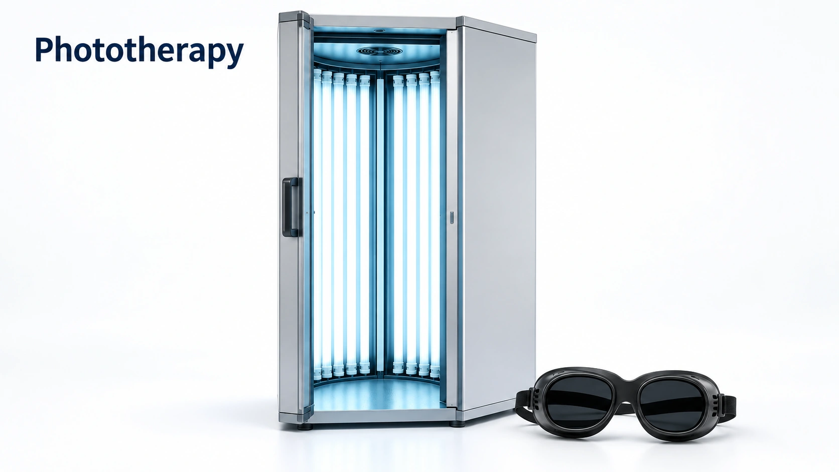
Phototherapy: A prescribed medical light treatment that uses a narrow band of ultraviolet B, usually two or three times a week, to calm psoriasis and eczema and to bring color back in vitiligo. A tanning bed is a different thing and is not a substitute.
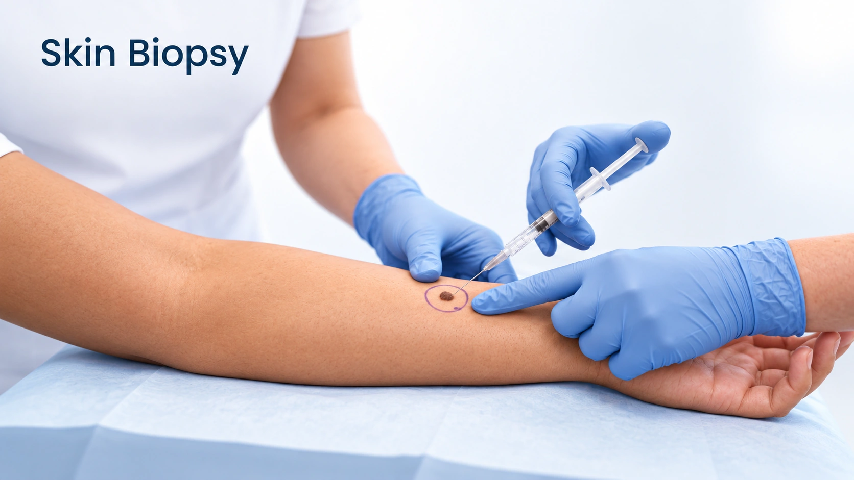
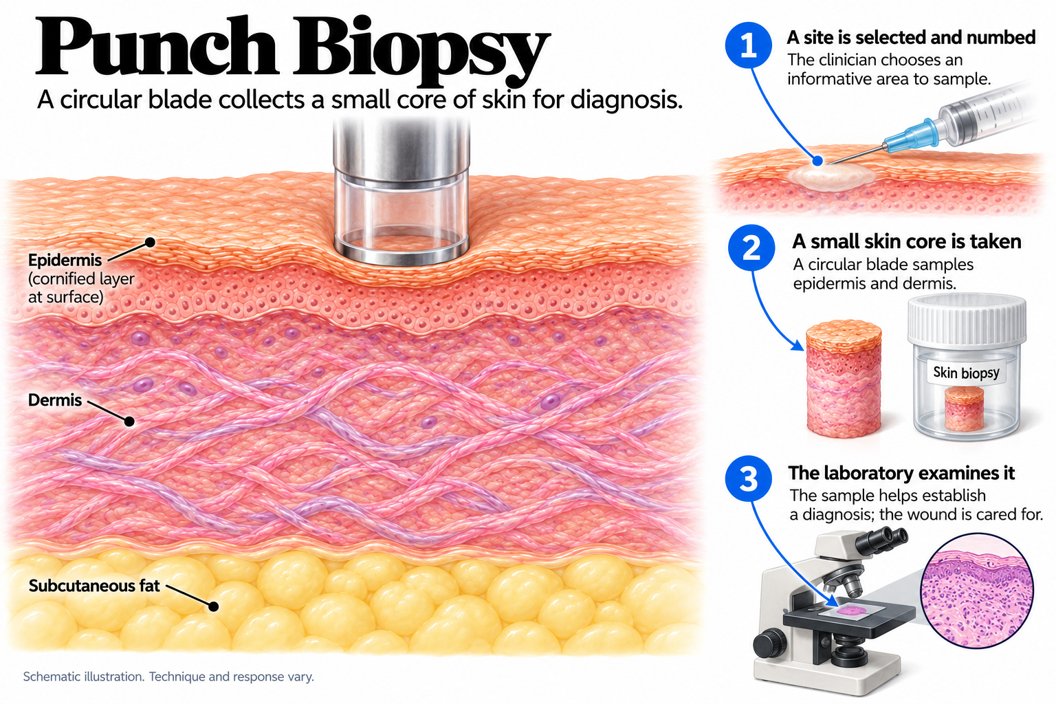
Punch Biopsy: A small round blade takes a full-thickness core of skin, usually 3 or 4 millimeters across, closed with a stitch or two. It is the standard biopsy for rashes and for anything where the deeper layers matter.
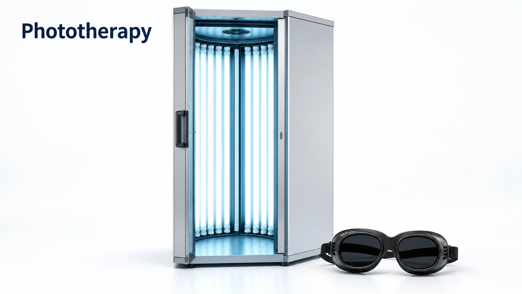

Choose for widespread psoriasis, eczema or vitiligo when creams can't cover enough skin and you can attend 2–3 times a week.
Choose when a rash or growth needs a definite diagnosis that looking alone can't give.
Can you have both?
They serve different purposes. A biopsy confirms the diagnosis; phototherapy treats conditions like psoriasis or eczema.
Controlled UV exposure treats selected inflammatory skin diseases.
PUVA combines a light-sensitizing medicine with UVA.
They are not equivalent to medical phototherapy.
Medicines, skin cancer history and cumulative exposure are checked.
A circular blade removes a small core of tissue.
It samples the epidermis, dermis and sometimes fat.
It is a diagnostic procedure.
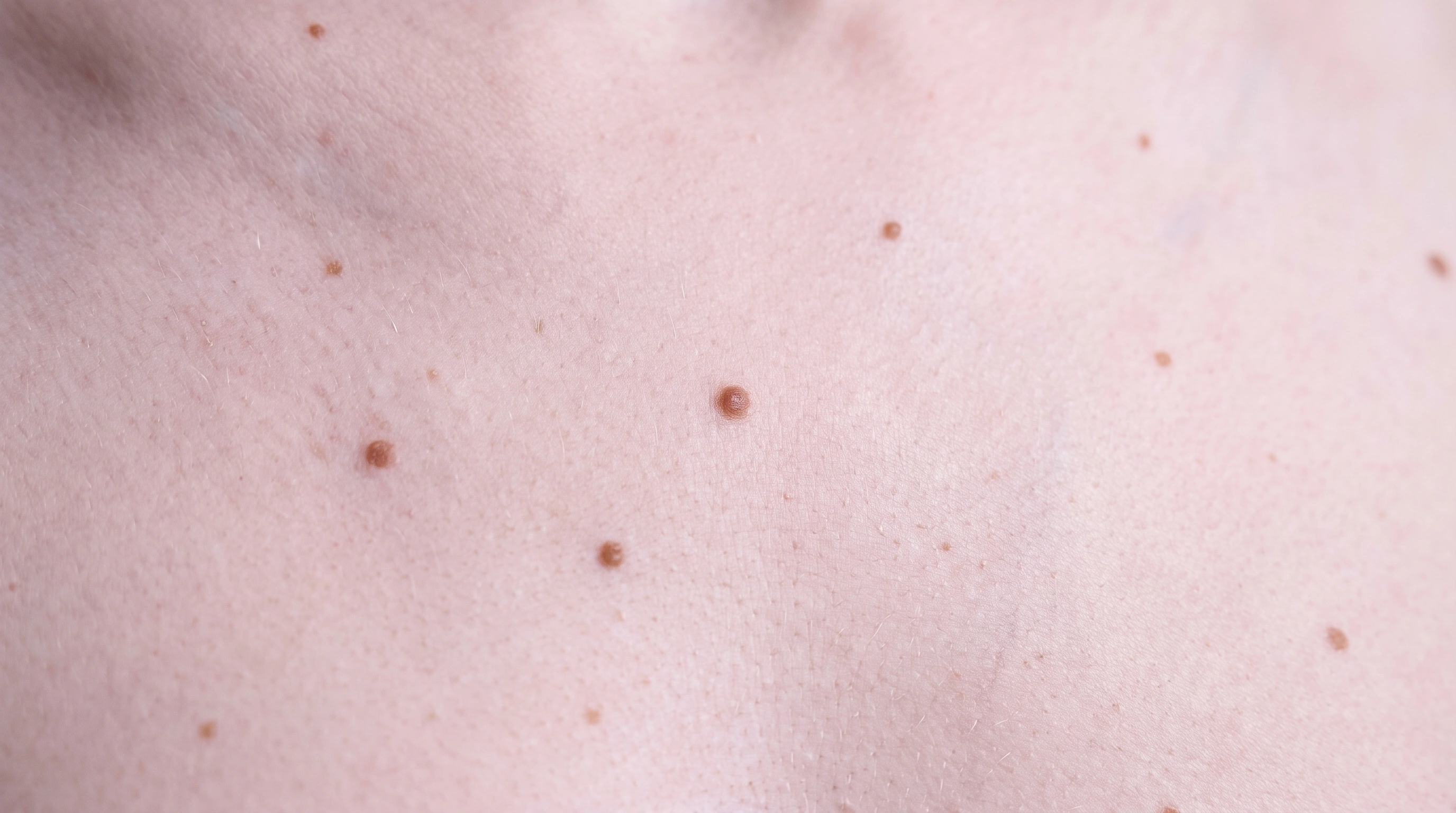
A partial sample may not characterize a whole mole.

Clears widespread plaques.
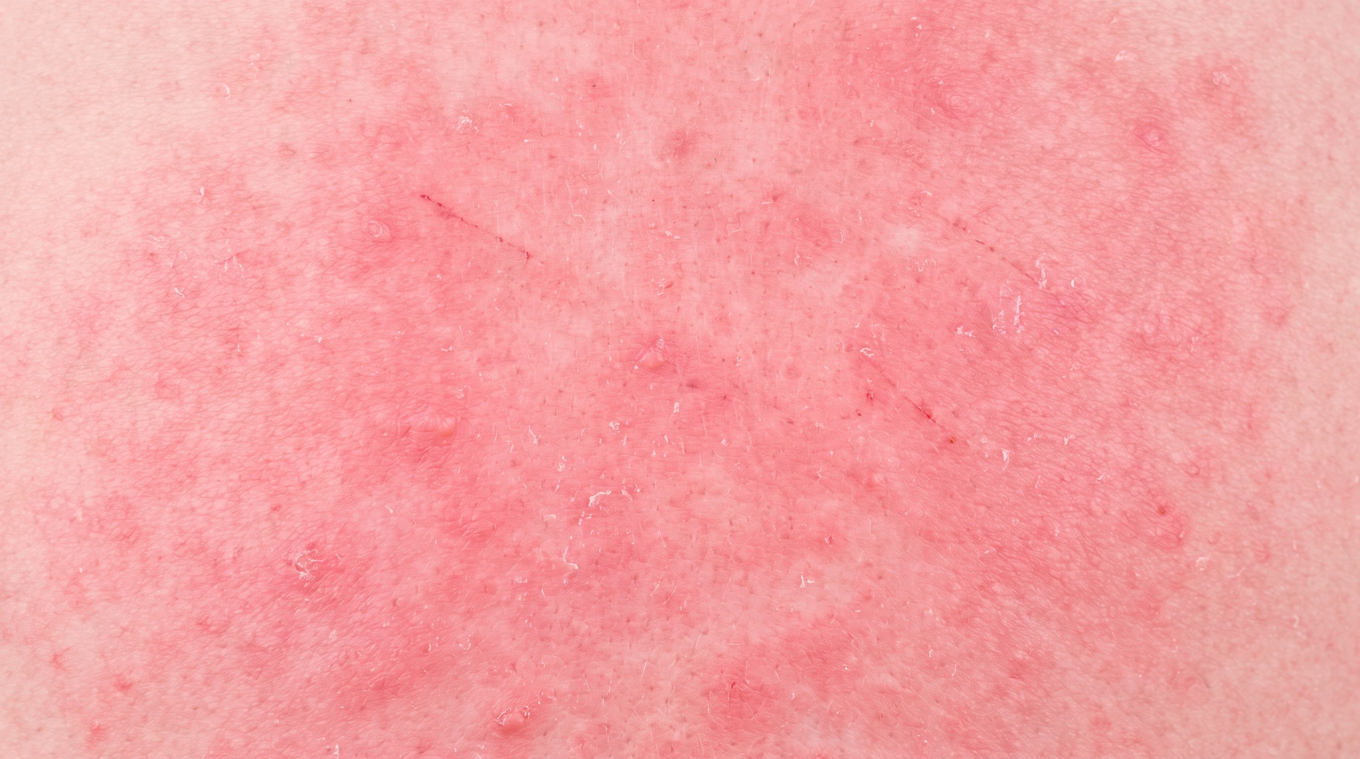
Reduces itch and rash.
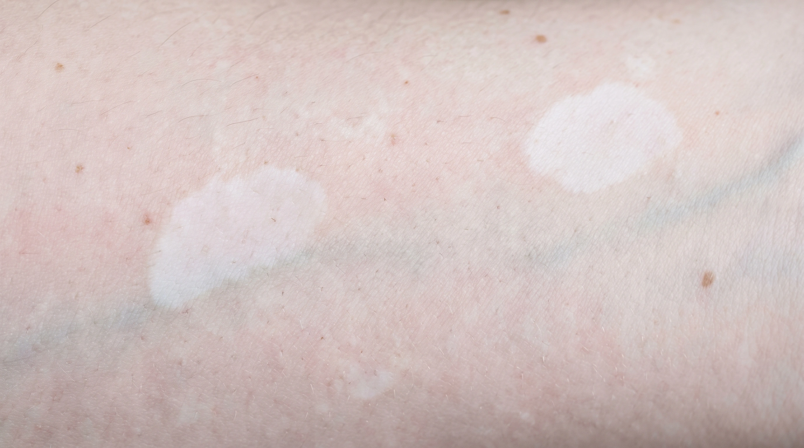
Restores color.
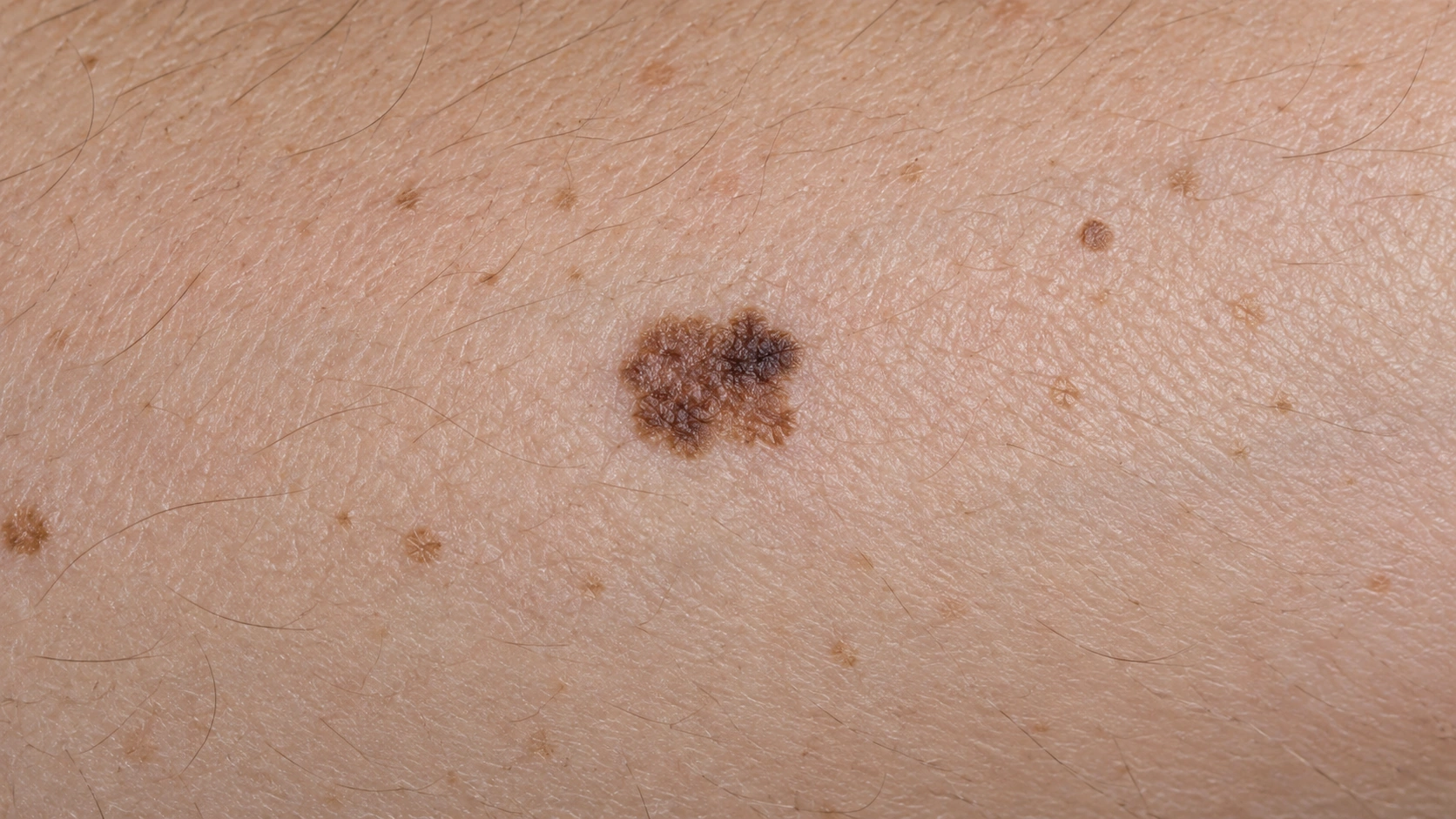
Confirms a diagnosis.
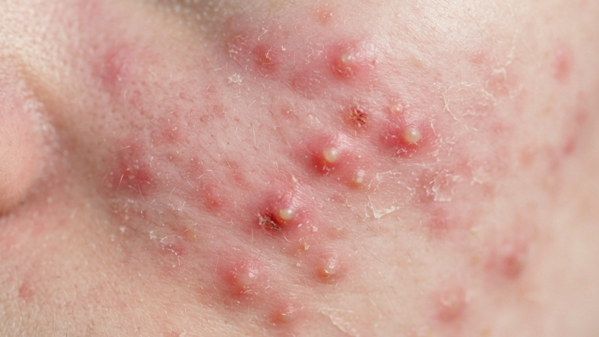
Confirms an unclear rash.
Not needed for diagnosis.
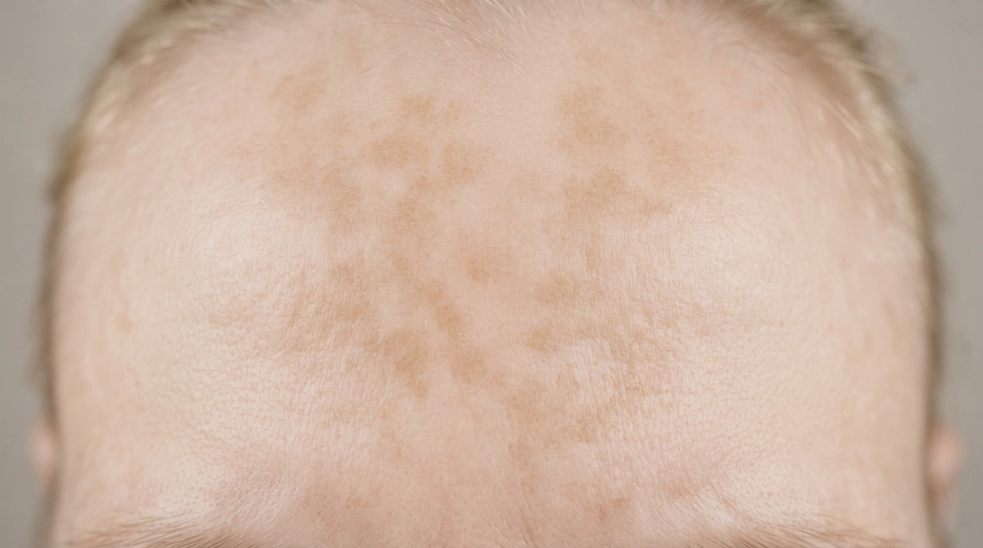
UV light darkens patches.
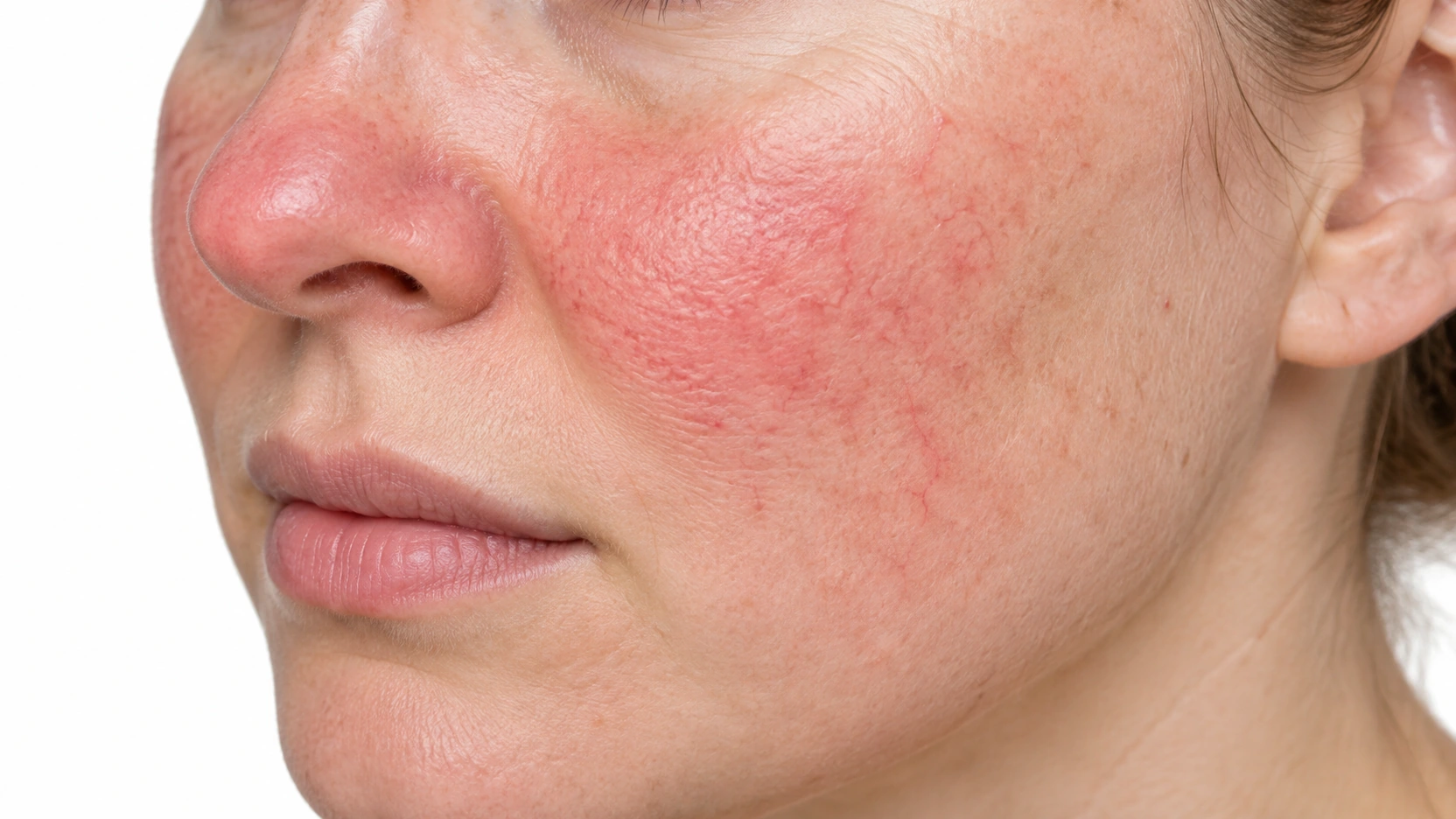
Light triggers flushing.
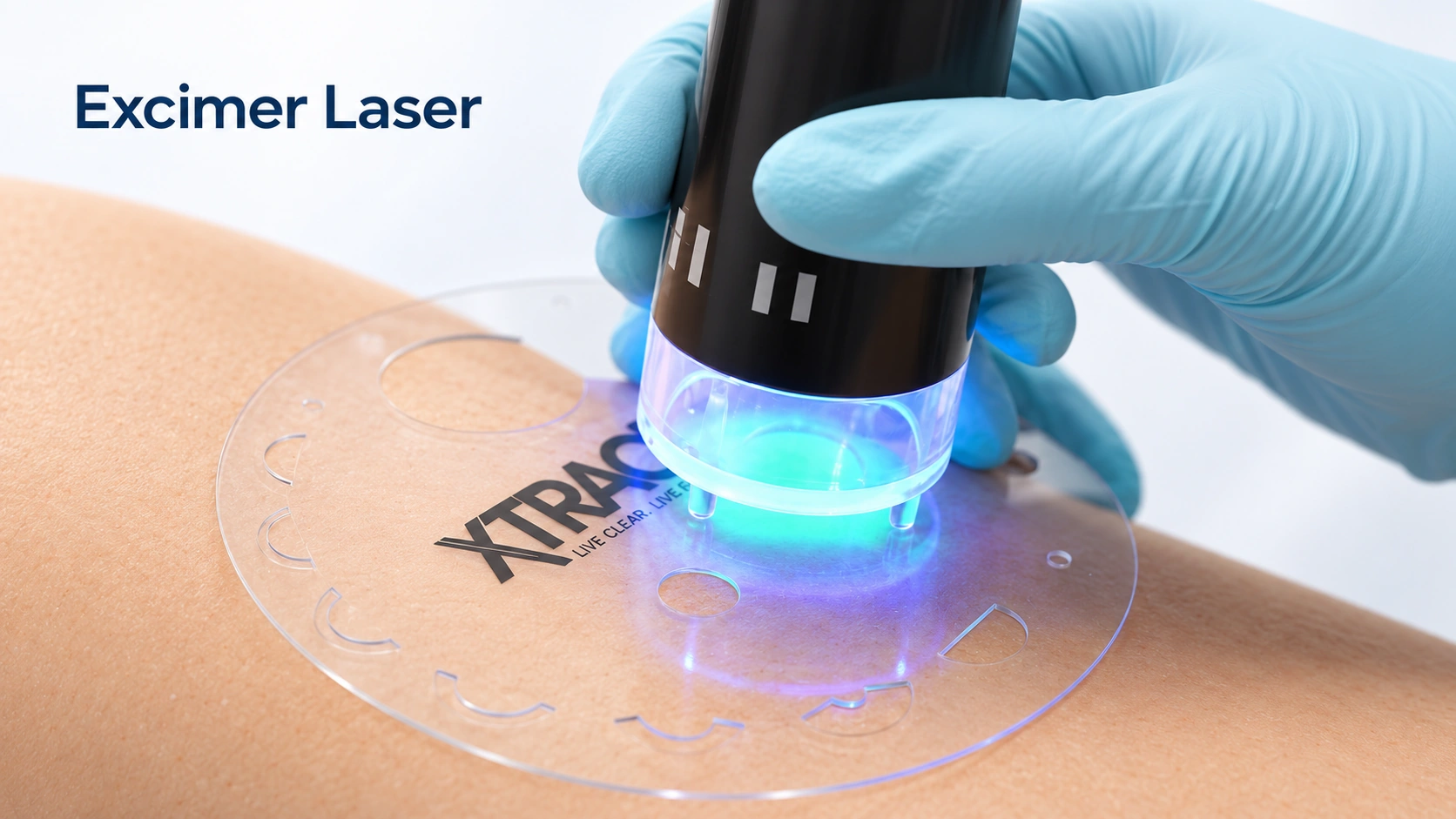
Affected skin may be treated in a whole-body booth or with a smaller unit; unaffected areas may be shielded.
The specific lesion, scar or affected patch determines the treatment site; there is no fixed body distribution.

Measured UV light reaches skin: A clinician selects and monitors the light exposure. Inflammatory activity changes: UV treatment can reduce overactive immune activity in skin. Skin symptoms may improve: Repeated treatment can help selected inflammatory conditions.
Schematic illustration. Technique and response vary. This explains a mechanism and does not specify treatment settings.
A site is selected and numbed: The clinician chooses an informative area to sample. A small skin core is taken: A circular blade samples epidermis and dermis. The laboratory examines it: The sample helps establish a diagnosis; the wound is cared for.
Schematic illustration. Technique and response vary. This explains a mechanism and does not specify treatment settings.
1 to 2 weeks before: Steroid cream: Stop the steroid cream a week or two before if you can.
Between sessions: Sun and tanning: No sunbathing, no tanning beds.
Clean skin: Emollient on plaques only if told to.
Goggles and shielding: Know which areas will be shielded, and bring your own goggles if you have them.
Eating and clothes: On the scalp a small patch is trimmed short, not shaved bald.
Eating and clothes: Eat normally, take your usual medicines, wear clothes that make the site easy to reach.
You are asked whether your skin burned, stung or went pink after the last session.
The site is chosen, and not always the one you would pick — for a rash, usually fresh untreated skin rather than the oldest patch.
You undress to the treated areas, put on goggles, and step into the cabinet.
Men get genital shielding — standard, not optional. The face is covered if it is not affected. Lamps run from a few seconds early in a course to several minutes later. You stand still and turn if asked. It does not hurt, and most people feel only warmth.
The skin is cleaned and stretched so the hole closes as a line, not a circle.
The punch is pressed and rotated, the core lifted out and cut free, then pressure stops the bleeding. One or two stitches and a dressing follow. You feel firm pressure and twisting, nothing sharp — say so if you do, and more anesthetic goes in.
Skin may look slightly pink and feel warm, like light sun.
A dressing for 24 hours, aftercare instructions, a stitch removal date, and a plan for how the result reaches you.
Days 1 to 2: A faint pink that fades by the next day is expected, and it is how the dose is judged.
Days 1 to 3: From day two, wash with soap and water once a day, pat dry, apply plain petroleum jelly and re-cover.
Days 5 to 14: Face stitches come out at about 5 to 7 days, elsewhere at 10 to 14.
Weeks 2 onward: The line is pink or purple and can feel firm or lumpy.
3–4 weeks to start
Lab report in 1–2 weeks
Months; vitiligo color can be lasting
One-time test
Treated skin dries out and tans.
Gaps matter.
Written estimate: Ask whether the quoted fee includes assessment, anesthesia, supplies, pathology when relevant, follow-up, and any planned repeat treatments.
In the US a punch biopsy usually produces two bills: the office fee, and a separate laboratory fee for reading the sample.
This is one of the few treatments here that insurance often pays for, because it treats a medical condition.
Insurance normally covers it when there is a medical reason, subject to your deductible and copay.
No. Tanning beds emit mostly ultraviolet A with a little UVB; this uses a narrow band around 311 nanometers chosen to keep the benefit and cut the burning wavelengths. Phototherapy doses are prescribed, recorded and adjusted, and areas that do not need treating are shielded. Tanning beds carry an established skin cancer risk.
Psoriasis usually starts improving by the third or fourth week and often clears over roughly 20 to 36 sessions. Eczema is slower, six to twelve weeks, with itch settling before the rash. Vitiligo cannot be judged before three months and often takes six to twelve months.
Two or three times a week for most of a course. This is the hardest part, and the most common reason people stop, so be honest with yourself before starting and ask about a home unit if it will not work.
PUVA, the older tablet-plus-UVA treatment, clearly raises risk at high cumulative doses. Narrowband UVB studies have not shown a clear increase, but long-term data are limited, which is why lifetime sessions are counted, unaffected skin is shielded, and skin checks continue.
Yes, with a prescription. Home units use the same lamps and dosing protocol, your dermatologist writes the schedule and reviews progress, and insurance sometimes pays because a unit costs less than a long series of office visits.
Psoriasis and eczema usually return over weeks to months, and the length of the break varies a lot. Some people go onto a lower maintenance frequency instead of stopping. Pigment regained in vitiligo is often kept, though it can be lost if the condition is still active.
Usually 3 or 4 millimeters across — smaller than a pencil eraser. The skin is stretched first, so the round hole closes as a short straight line.
The numbing injection stings for a few seconds. After that you feel firm pressure and twisting, nothing sharp. Most people take nothing stronger than acetaminophen, on the first evening only.
Usually one or two. On the face they come out at about 5 to 7 days, elsewhere at 10 to 14. Absorbable stitches dissolve on their own and are an option if getting back is hard.
For rashes, fresh untreated skin shows the diagnostic changes clearly, while the oldest patch has scratched, treated or healed skin over it. The site is chosen for what the pathologist needs to see.
Normal for rashes. The report describes a pattern that narrows the list, and the doctor combines it with what your skin looks like. Sometimes a second biopsy at a different stage settles it.
Yes — a small line or dot, a few millimeters. It stays pink for a couple of months, then fades. On deeper skin tones a brown mark is common for months, and raised scars are more likely on the chest, shoulders and upper back.