It is a clinical procedure.
Moles are removed for diagnosis, symptoms or cosmetic reasons.
Mole Removal: Taking a mole off, either because it looks unusual or because you dislike it. The method matters less than one rule: a pigmented spot that is removed should be sent to the lab, because that is the only way anyone finds out what it was.
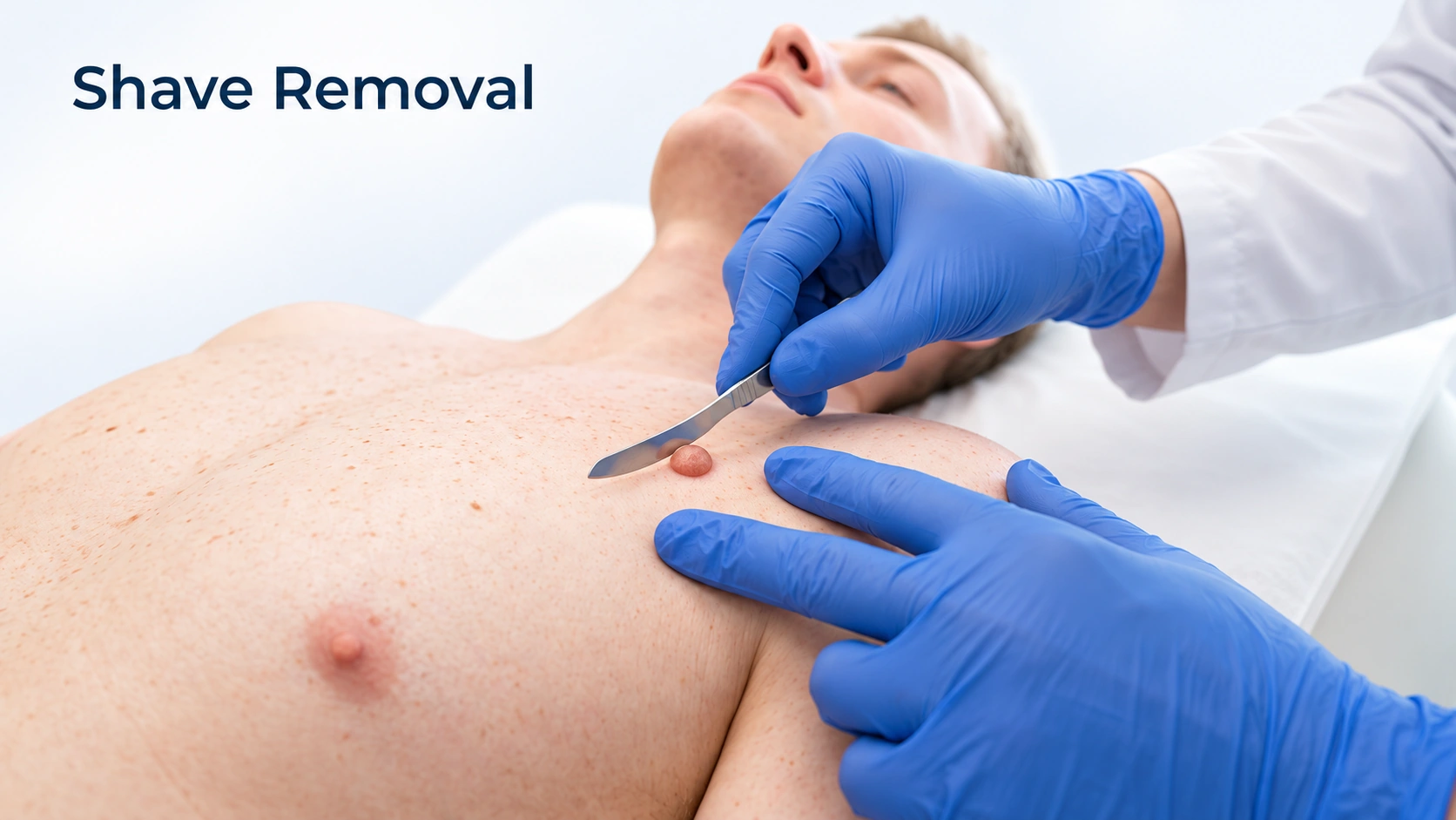
Shave Removal: A raised spot is numbed and shaved off level with the skin using a small flexible blade. No stitches and no hole — a scab that heals in a week or two. The same procedure can remove a harmless bump or take a sample for the laboratory.
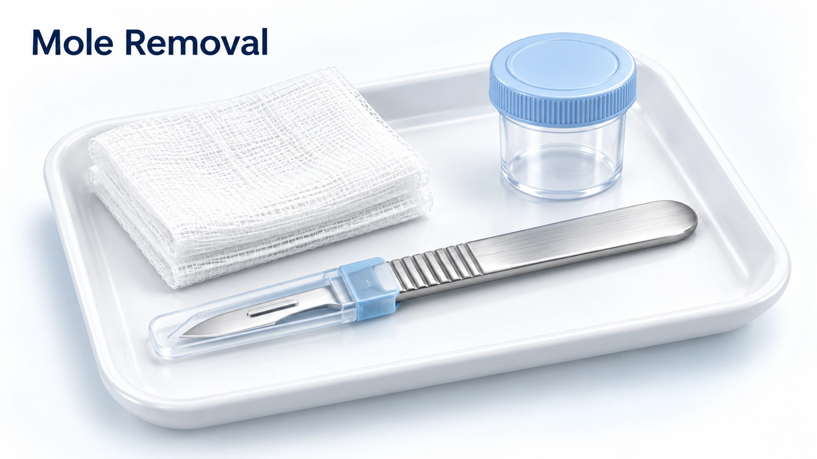
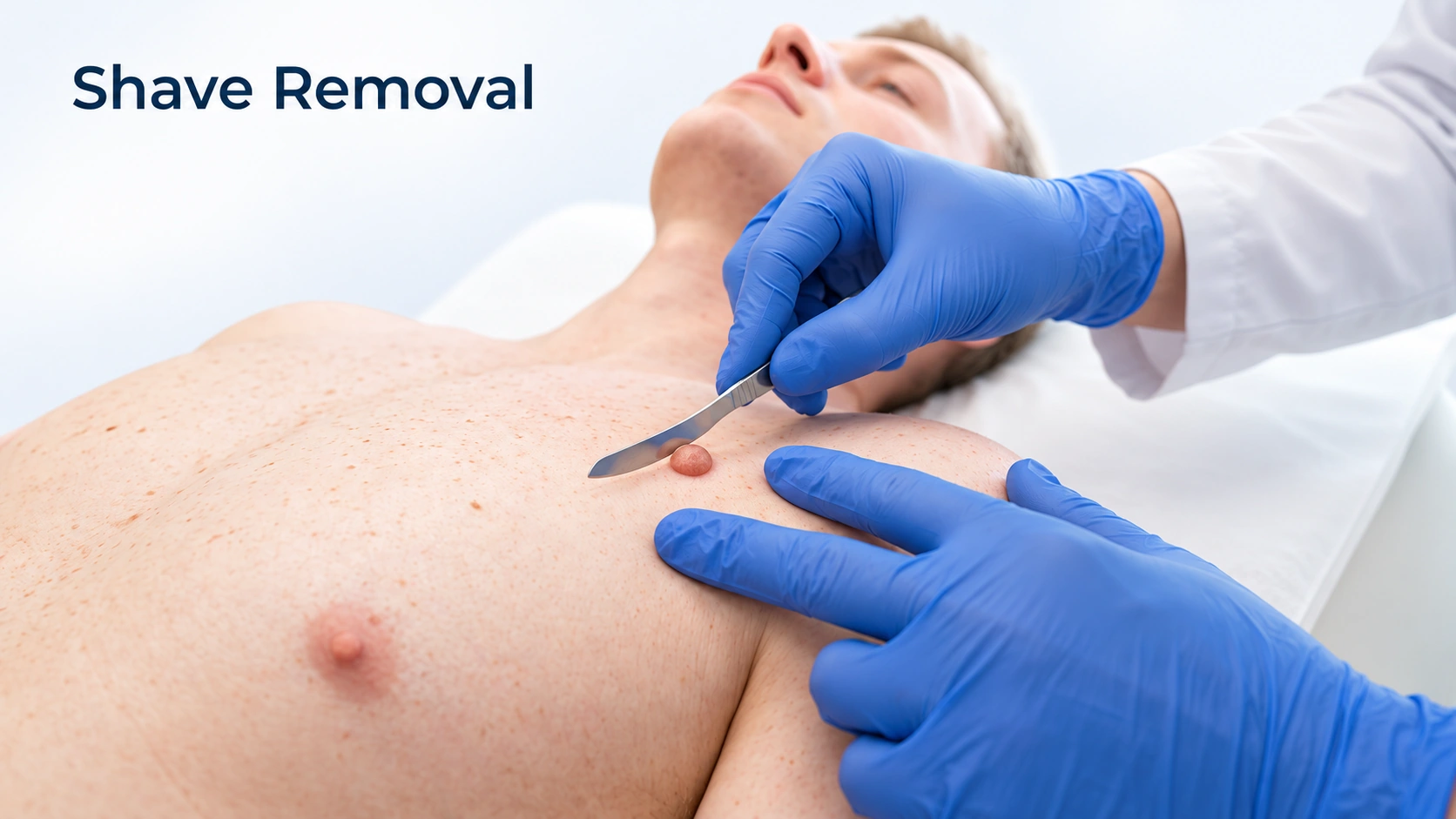
Choose to remove a mole that looks unusual or bothers you — and always have it sent to the lab.
Choose to remove a raised bump with no stitches — a flat mark that heals in a week or two.
Can you have both?
Shave removal is one way to remove a mole. Your dermatologist chooses the method for each mole.
Moles are removed for diagnosis, symptoms or cosmetic reasons.
A possible cancer needs a tissue specimen.
Acids and home devices can destroy diagnostic information.
Tissue must be preserved to assess depth.
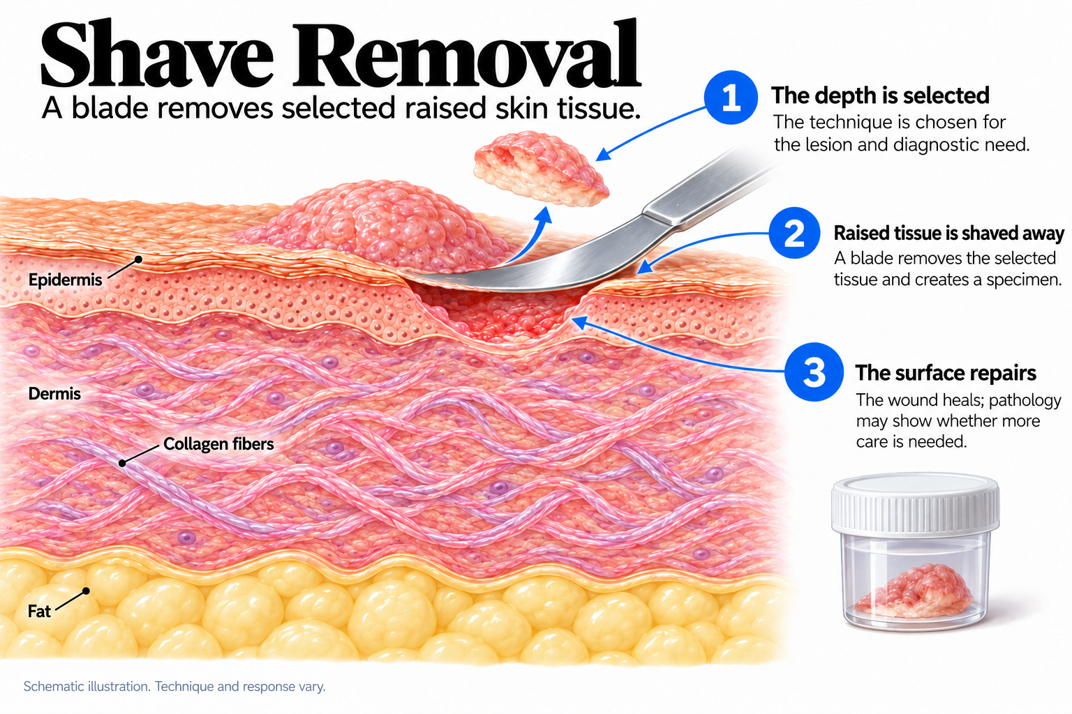
A lesion is shaved off at a chosen depth under local anesthesia.
It can remove raised benign growths or obtain a specimen.
The clinician chooses the technique.
Cosmetic removal must not replace adequate sampling.
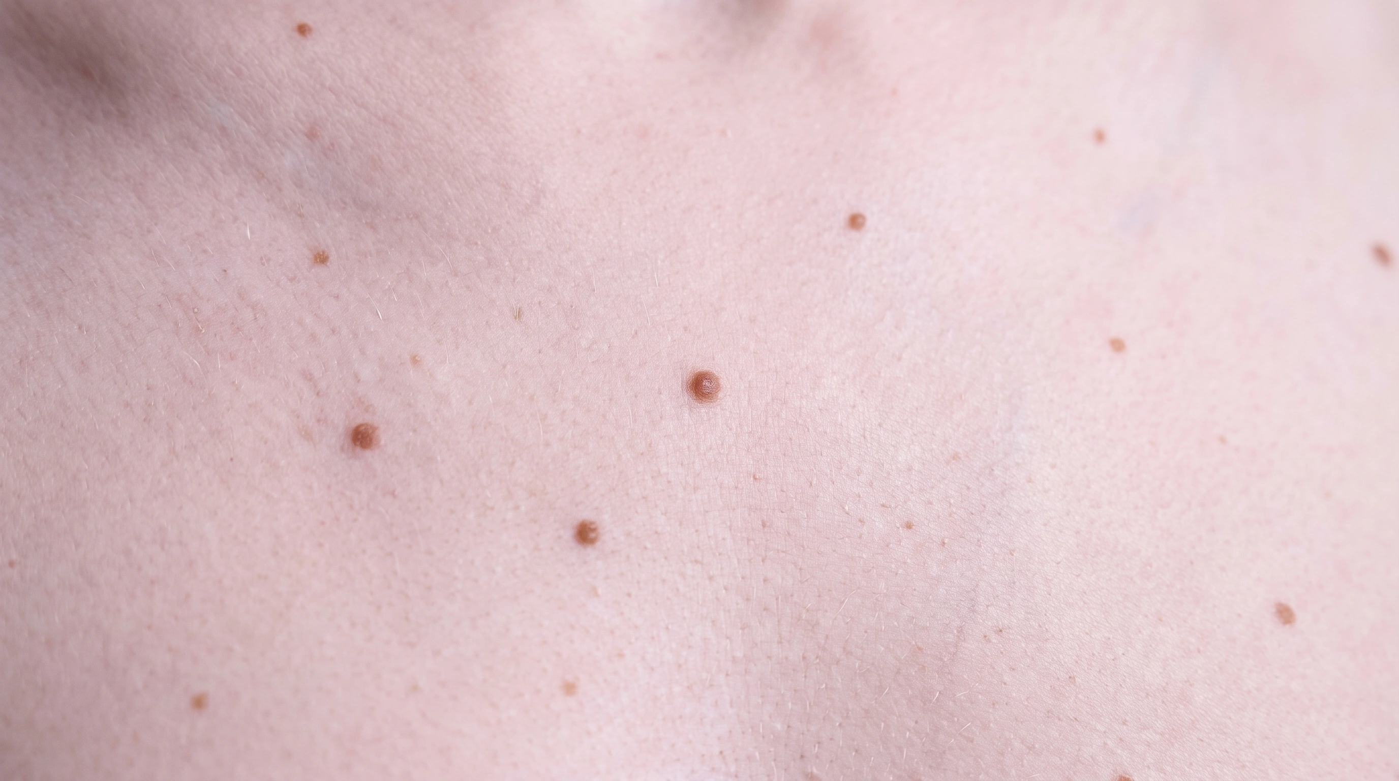
Removes the mole and checks it.
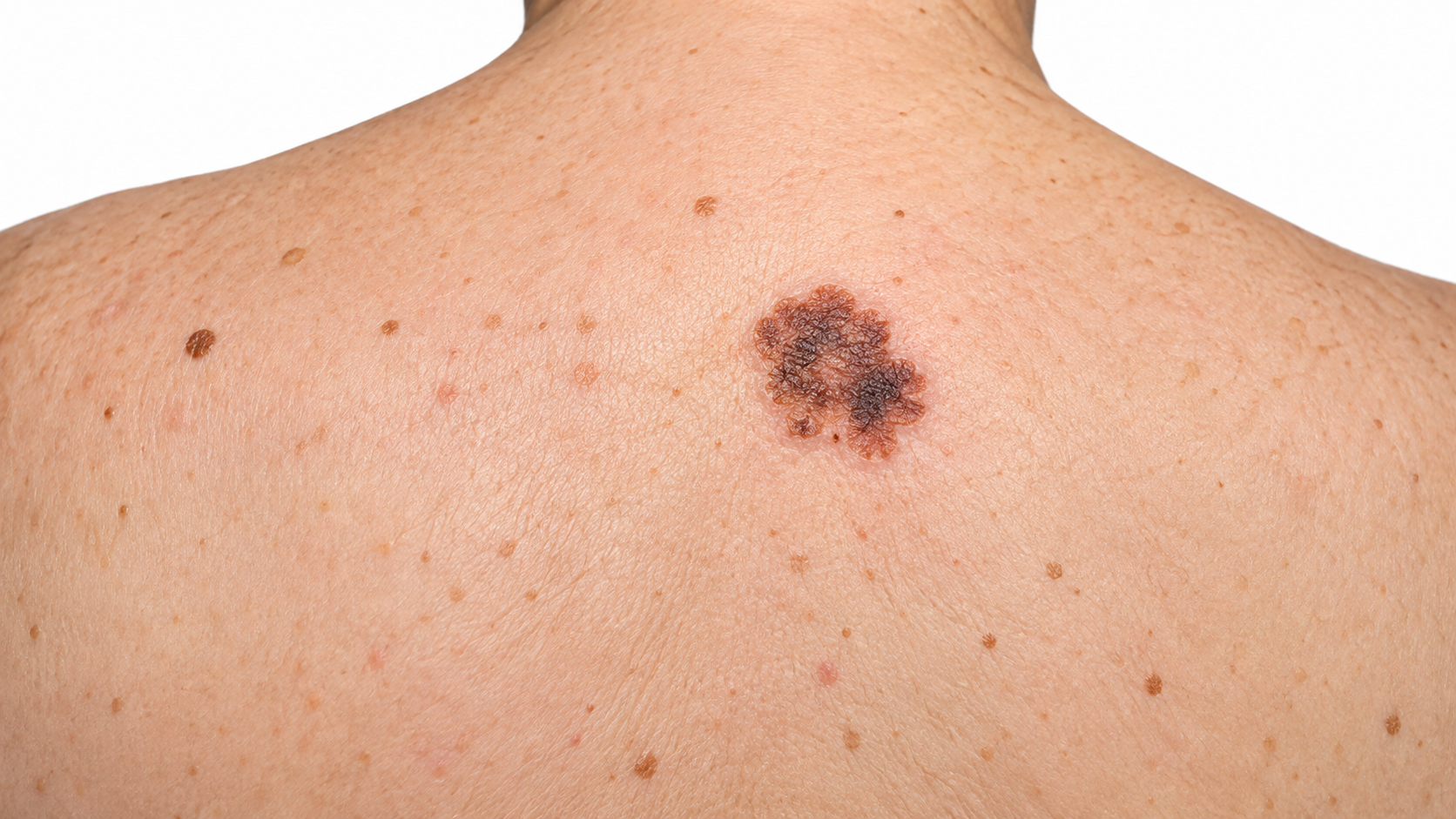
Excision is the first treatment.
Removes raised moles.
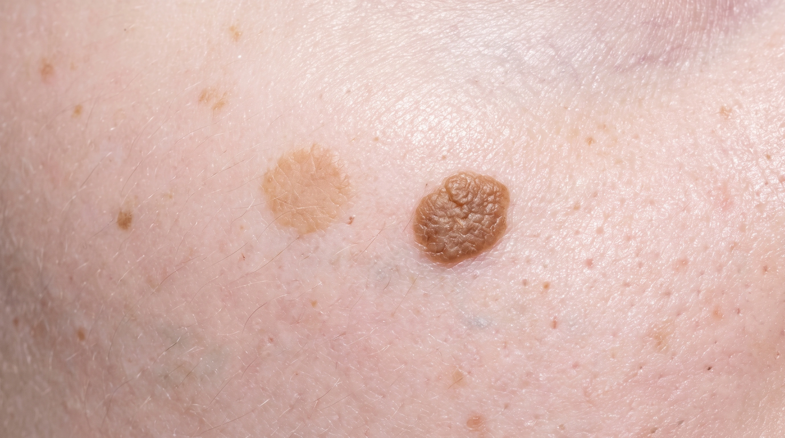
Removes raised growths.
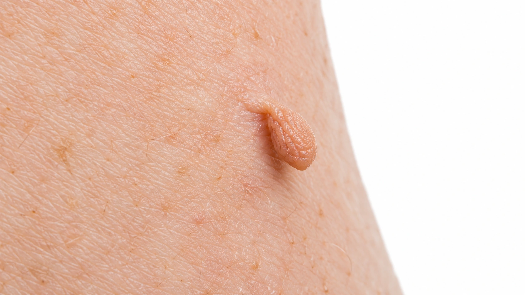
Quick removal.
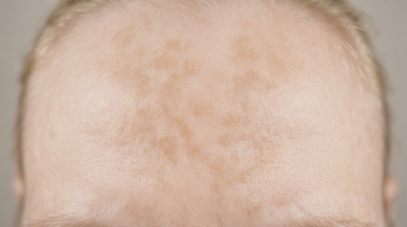
Not a mole.
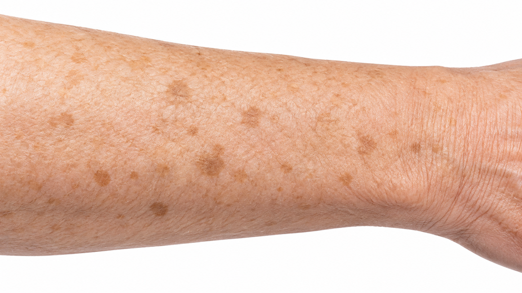
Lasers or creams fit flat spots better.
Needs full excision.
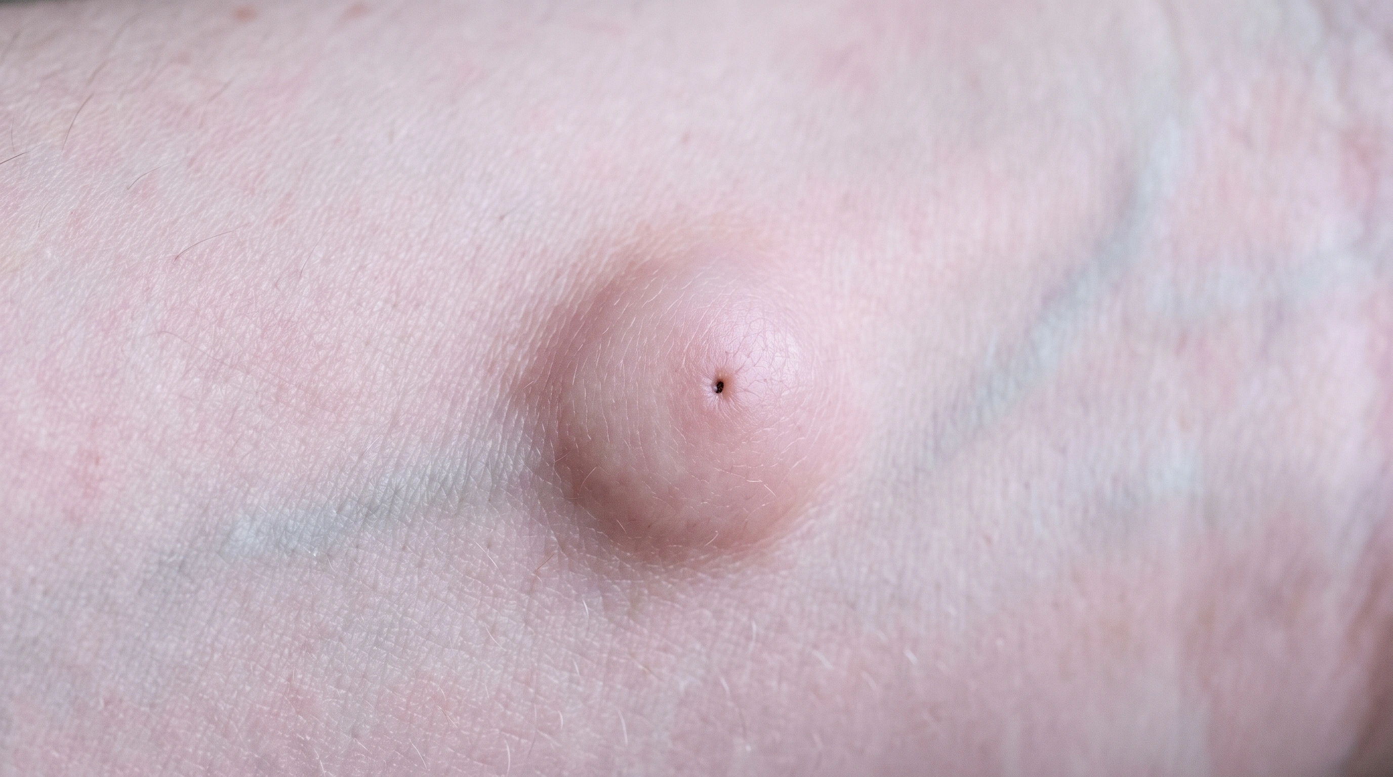
The sac stays and regrows.
The specific lesion, scar or affected patch determines the treatment site; there is no fixed body distribution.
The specific lesion, scar or affected patch determines the treatment site; there is no fixed body distribution.

The mole is assessed: Examination guides the technique and depth of removal. Selected tissue is removed: A clinician removes the mole with an appropriate method. Healing and review follow: A scar forms; laboratory findings may guide further care.
Schematic illustration. Technique and response vary. This explains a mechanism and does not specify treatment settings.
The depth is selected: The technique is chosen for the lesion and diagnostic need. Raised tissue is shaved away: A blade removes the selected tissue and creates a specimen. The surface repairs: The wound heals; pathology may show whether more care is needed.
Schematic illustration. Technique and response vary. This explains a mechanism and does not specify treatment settings.
Plan ahead: Lifting: On the back, shoulder or leg, expect a week or two of no heavy lifting or gym after stitches.
Photo and sun: Photograph the mole in good light beside something for scale.
What gets removed: Decide what you want done with each spot, and ask that anything removed goes to the laboratory.
Spots and the exam: Point out every spot you are worried about at the start, since the plan for one can depend on the others.
Eating and clothes: Eat normally, take your usual medicines, and wear clothes that give easy access.
The mole is examined with a dermatoscope and often photographed.
The spot is examined, often with a handheld magnifier, and measured or photographed.
For a shave, a fine blade is drawn flat across the base and the site sealed with a solution or light cautery — no stitches, a couple of minutes.
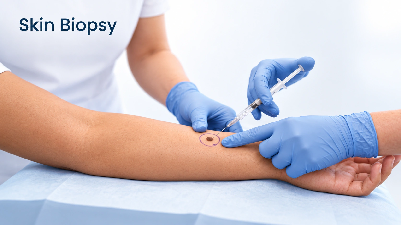
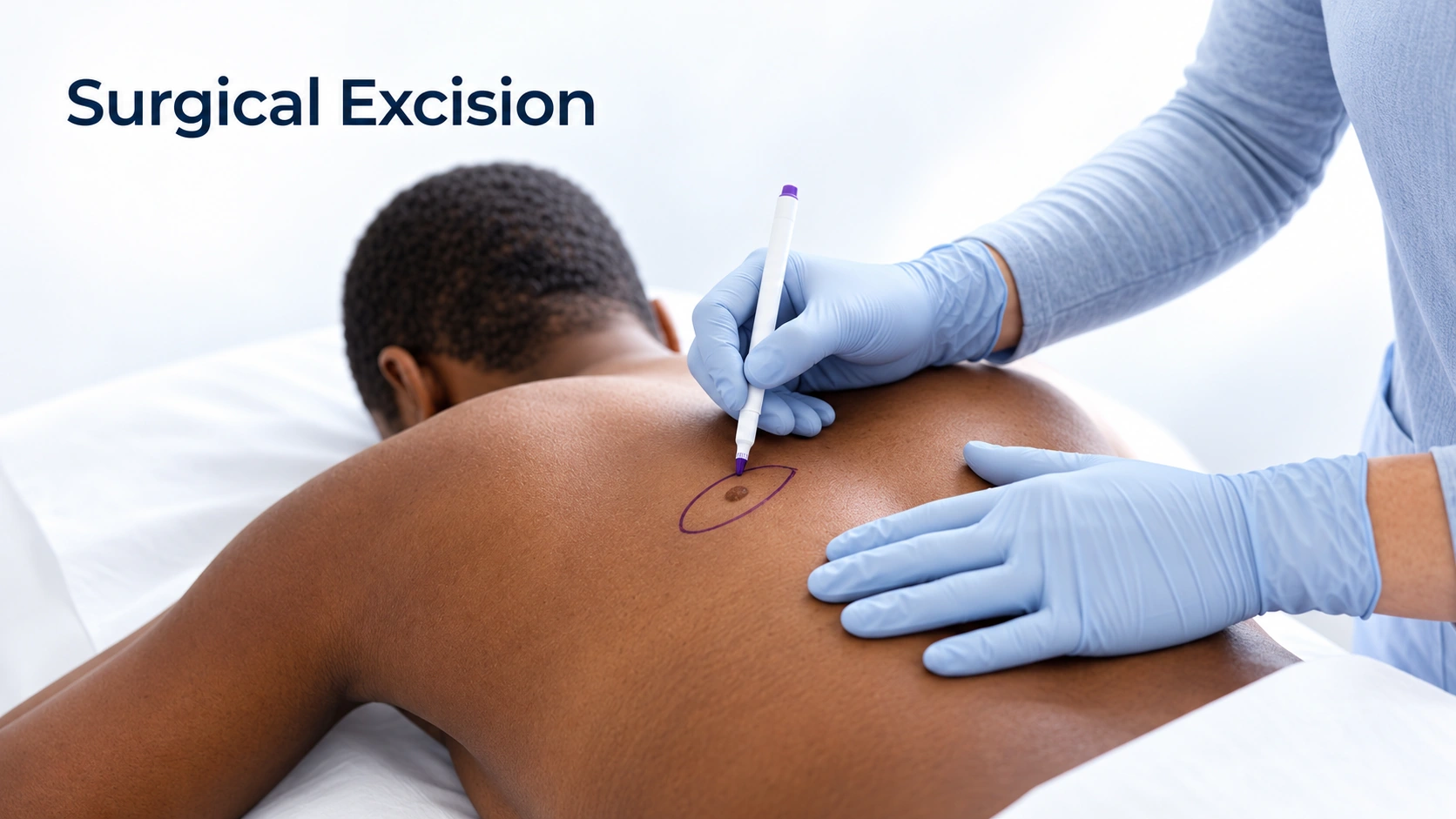
A punch or excision takes 15 to 30 minutes: the skin is cut, the specimen lifted out, the wound stitched. You are awake and feel pressure and tugging, not sharpness. Anything sharp means you need more anesthetic.
The skin is cleaned, and a blade held nearly parallel to the surface slices the lesion off in seconds.
Bleeding is stopped with a solution on a cotton bud or light cautery — it can smell faintly of burning and is not painful. You feel pressure and scraping, not cutting; say something if anything feels sharp.
The specimen goes into a labeled pot in front of you and a dressing goes on.
A dressing, aftercare instructions, and a plan for how the result reaches you.
Days 1 to 14: Keep a shave site moist under petrolatum and covered rather than letting it dry into a hard scab; a little ooze early on is normal.
Days 1 to 3: The anesthetic wears off over one to three hours, and the site is tender and may ooze.
Days 4 to 14: New skin fills the site and any scab lifts on its own.
Weeks 1 to 2: Most lab reports come back now.
From week 2: A flat pink circle sits where the spot was, looking more obvious than the final result.
Months 1 to 12: A shave site stays a flat pink or pale circle that fades slowly.
Immediately; lab report in 1–2 weeks
Immediately
Permanent if fully removed
Usually permanent; some moles regrow pigment
In the US, a single mole removal in a dermatology office commonly costs a few hundred dollars — more for an excision than a shave, and more on the face.
Removal of skin tags and keratoses sits on the line.
A mole removed because it looks concerning is a diagnostic procedure, and insurance usually covers it, subject to your deductible and copay.
In the US, cost depends on why it was done.
If it is pigmented, yes, and it is worth insisting on. Testing is the only way to know what it was, it costs a separate lab fee rather than another procedure, and the alternative is a permanent gap in your record. If a provider proposes removing a mole without sending it, ask why.
No. There is no evidence for it, and removal with testing is exactly how melanoma is diagnosed and, caught early, cured. The delay this belief causes is the actual harm.
After a shave, some color can return because deeper pigment cells were left behind. It is usually harmless, but it always gets examined and often means another biopsy to be certain. Full-thickness removals rarely regrow.
Yes, some mark is unavoidable. A shave leaves a flat pale or pink circle about the width of the mole. An excision leaves a line longer than the mole was, because the skin has to be closed neatly. On the face, discuss that trade before the day.
Yes. Cosmetic removal is a legitimate and common reason, and the requirements do not change: examined first, an appropriate method, tissue sent to the lab. It is usually self-pay.
Usually one to two weeks. Call if nothing arrives by then, and assume nothing from silence — missed results are an administrative problem, not a sign that everything was normal. Ask for a copy of the report either way.
No. It takes off the raised part and leaves a shallow wound level with the skin, which fills in over one to two weeks. The usual result is a flat mark, slightly paler than the skin around it.
It can. Keratoses and skin tags sometimes regrow from the base, and a shaved mole can return with color months later because pigment cells survived underneath. Have regrowth looked at, and mention that the spot was shaved.
Usually not. A shave normally samples a basal cell or squamous cell cancer rather than clearing it, and a second procedure follows. Keep that appointment even though the skin looks healed.
Often yes, if it has been examined and looks harmless. It is billed as cosmetic, and the trade is a flat pale mark instead of a raised bump. It should still go to the laboratory.
No. That is the main appeal — the wound heals from the base under a layer of petroleum jelly.
Keep the site moist with plain petroleum jelly and covered until it closes over, do not pick, and keep it out of the sun for several months. Drying out and sun are what turn a small shave site into a lasting mark.