Basal Cell Carcinoma
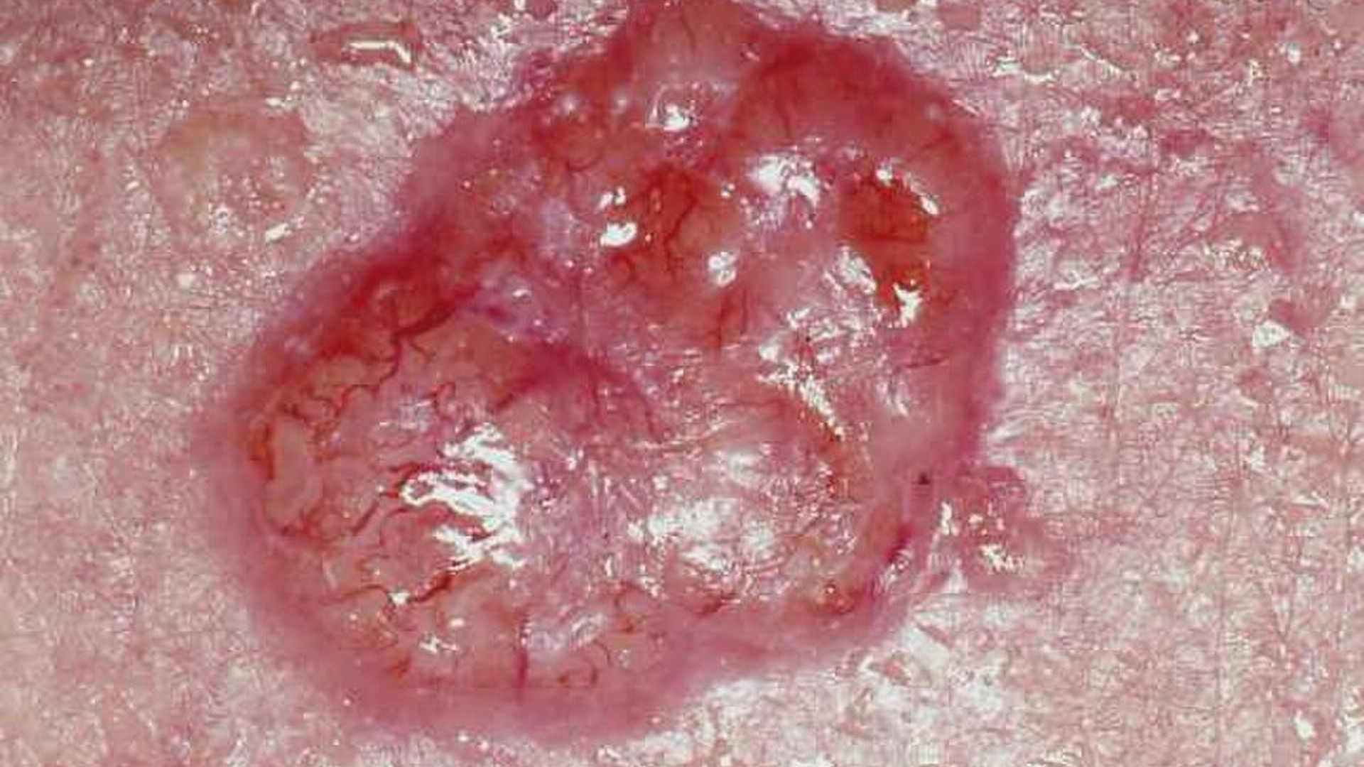
A basal cell carcinoma. The surface looks pearly and slightly see-through, with fine blood vessels running across it and a raised, rolled edge.
Basal cell carcinoma usually looks like a pearly or see-through bump, sometimes with tiny blood vessels across it. It may bleed, scab over, and never quite heal. Squamous cell carcinoma is more often a firm, scaly or crusted lump that can be tender and grows faster. Basal cell almost never spreads. Squamous cell has a greater risk of spreading. Both need prompt medical assessment and treatment.
| The difference | Basal Cell Carcinoma | Squamous Cell Carcinoma |
|---|---|---|
| What it looks like | Pearly, shiny, see-through | Rough, scaly, crusted |
| How it feels | Usually painless | Often tender |
| Treatment | Cut it out | Cut it out, and check further if needed |
A basal cell carcinoma. The surface looks pearly and slightly see-through, with fine blood vessels running across it and a raised, rolled edge.
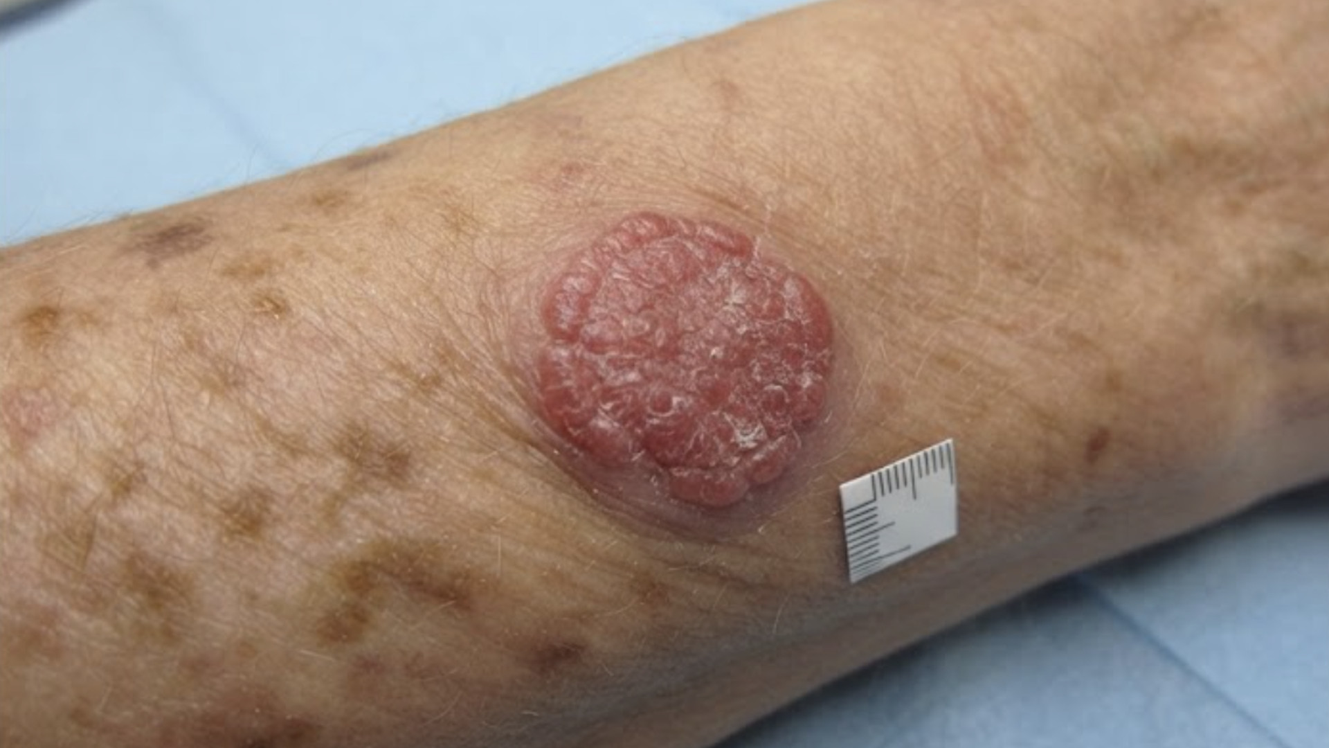
A squamous cell carcinoma. It is firmer and thicker, with a rough, scaly or crusted top instead of a pearly shine.
Side by side
Basal Cell Carcinoma
The most common cancer of any kind
About 80% of skin cancers that are not melanoma.
Squamous Cell Carcinoma
Second most common
About 20%.
Basal Cell Carcinoma
Pearly, shiny, see-through
A small raised bump with a glossy or waxy surface. Tiny blood vessels may run across it. It can also look like a pale flat scar, or a sore that will not heal.
Squamous Cell Carcinoma
Rough, scaly, crusted
A firm red bump, a scaly patch, or a sore with a raised, hard edge. It often has a thick crust that keeps coming back.
Basal Cell Carcinoma
Usually painless
It may bleed easily from minor contact, then scab, then bleed again.
Squamous Cell Carcinoma
Often tender
It is more likely to be sore or painful to touch, and more likely to feel firm and fixed.
Basal Cell Carcinoma
Slowly, over months to years
Squamous Cell Carcinoma
Faster, over weeks to months
Basal Cell Carcinoma
Sun-exposed skin
Face, nose, ears, scalp, neck, shoulders. The nose is the single most common spot.
Squamous Cell Carcinoma
Sun-exposed skin, plus some others
Face, ears, lips, backs of hands, forearms. Also on scars, chronic wounds, and inside the mouth or on the genitals.
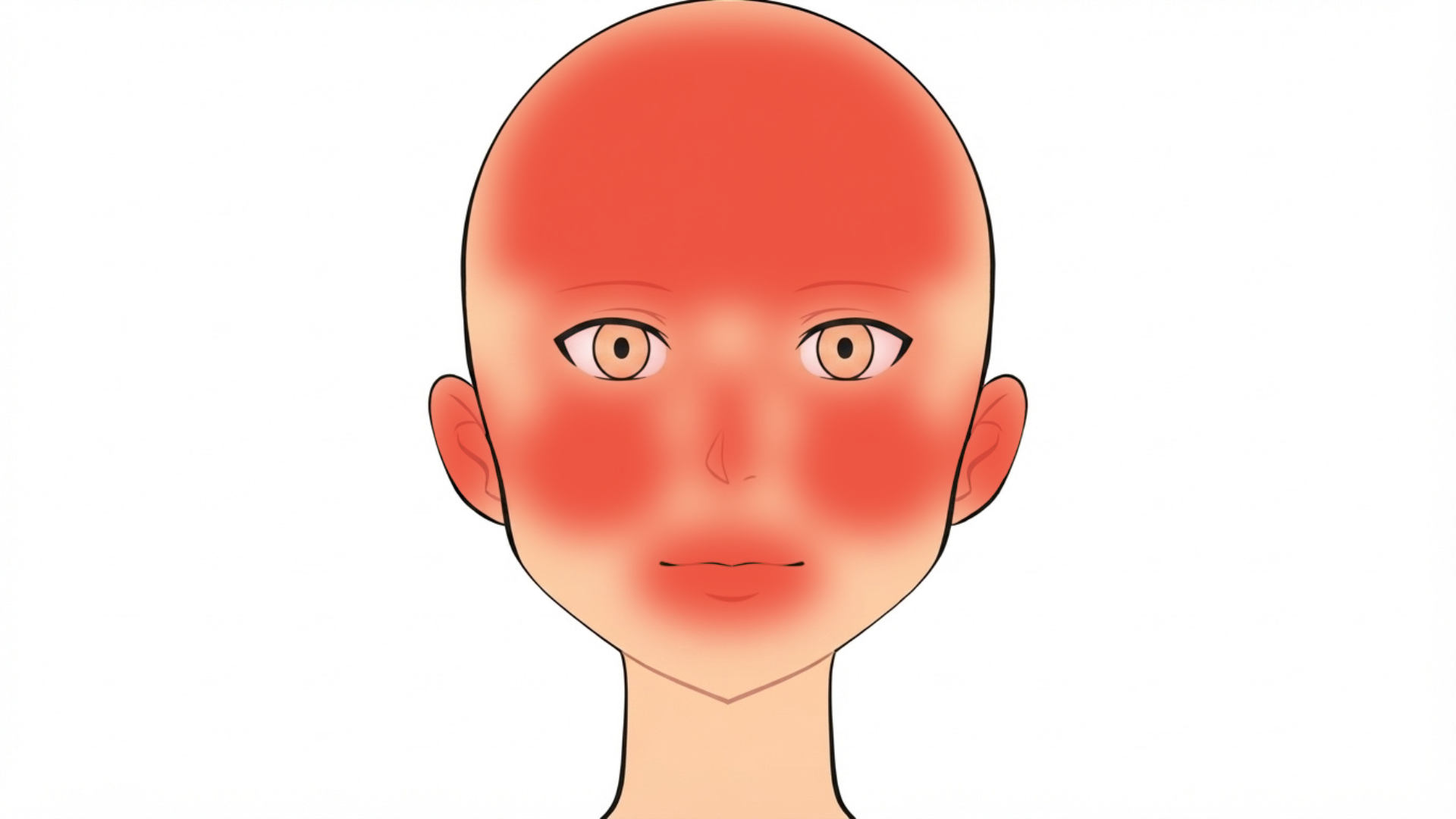
About eight in ten appear on the head and neck, because that is the skin exposed year-round. The nose is the single most common site, then the forehead, cheeks, ears, eyelids, lips, and the scalp in people with thinning hair.
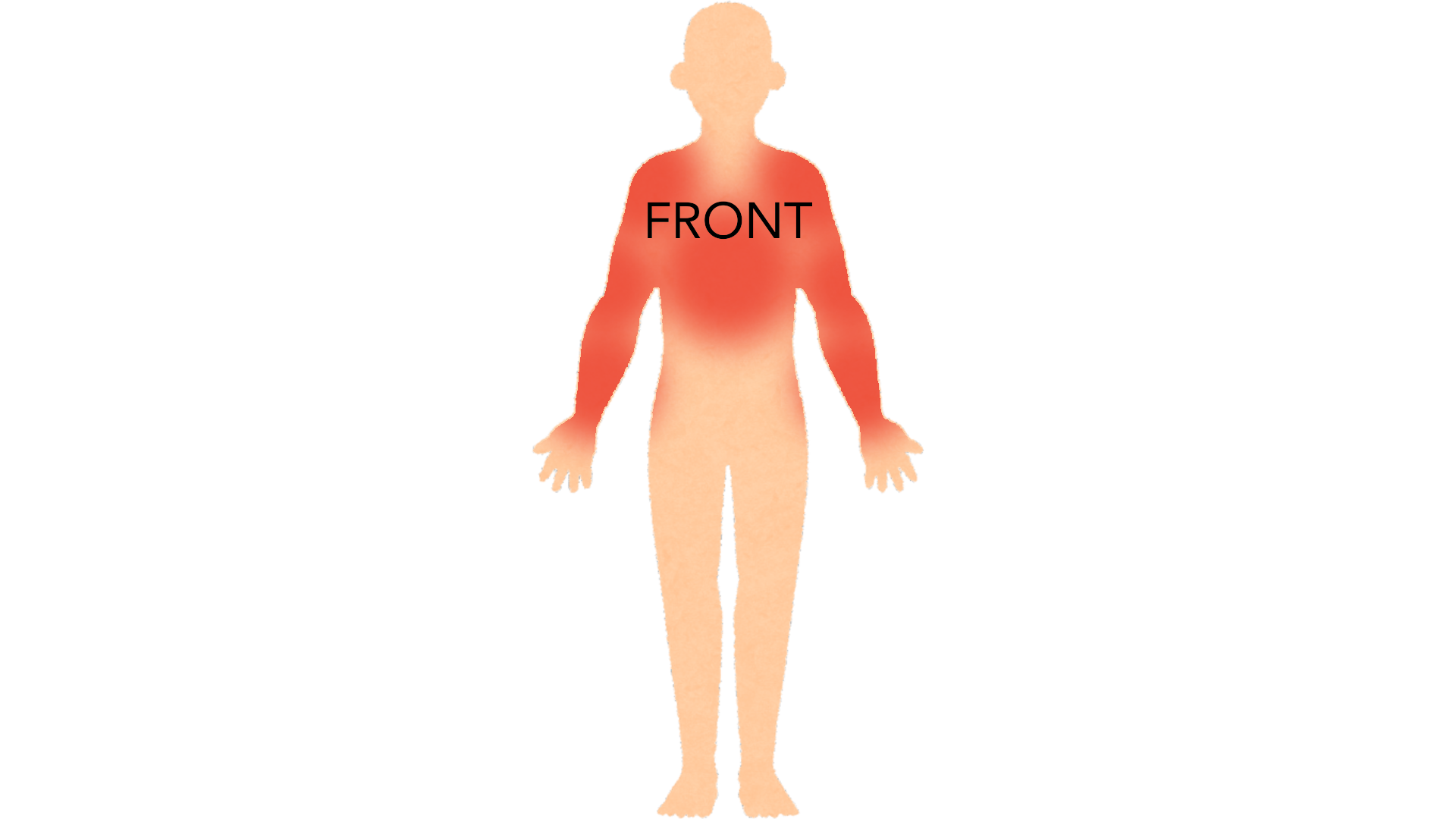
The rest appear on skin that gets sun in summer: the chest, the shoulders and the arms. The flat, scaly kind that spreads sideways rather than building into a lump is most often on the trunk.
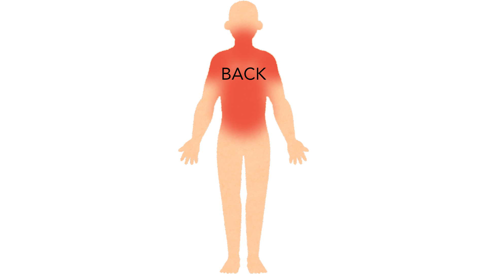
The back of the neck and the upper back are common in people who have worked or played outdoors, and they are the sites found late because nobody sees them. A sore on the back that scabs, heals and comes back in the same place is worth having looked at.
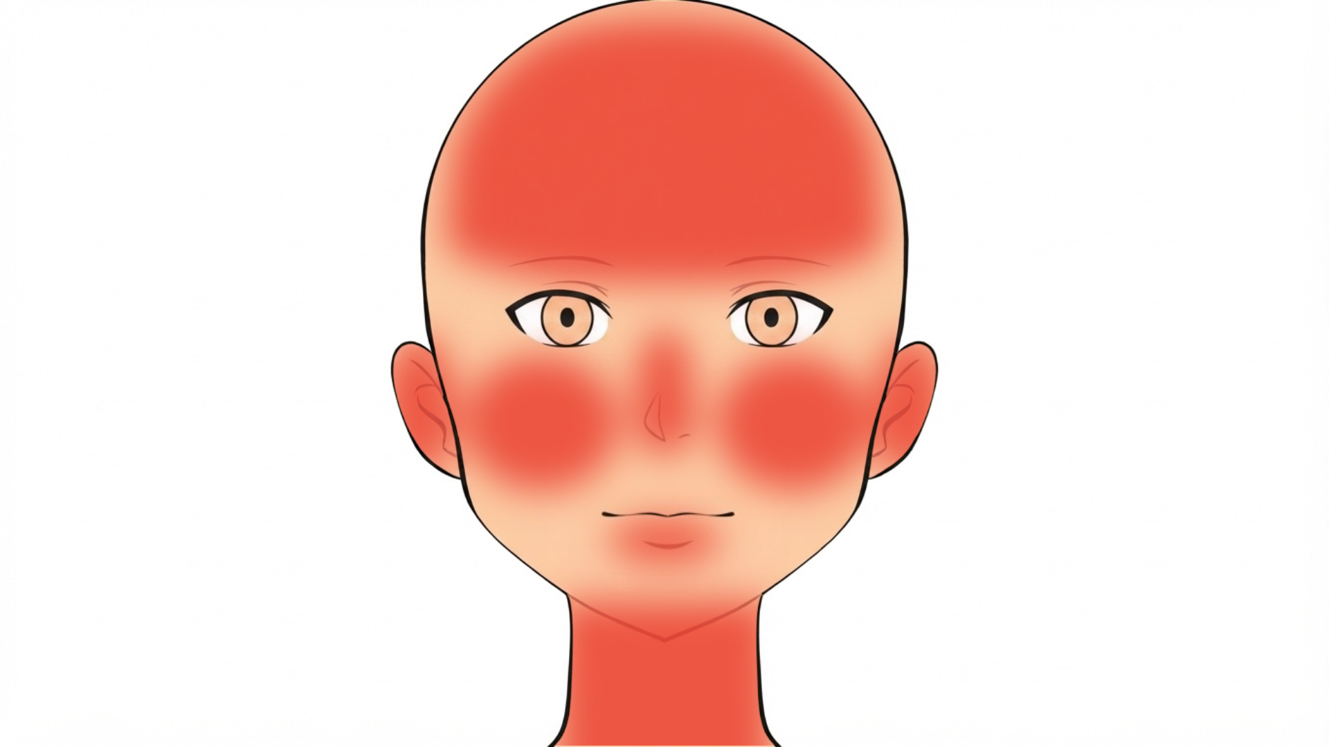
It turns up where sun has landed for decades: the face, the ears, the lower lip, the nose, a scalp with thinning hair and the neck. The ear and the lip are the two that matter most, because tumors there behave more aggressively than the same thing on a cheek.
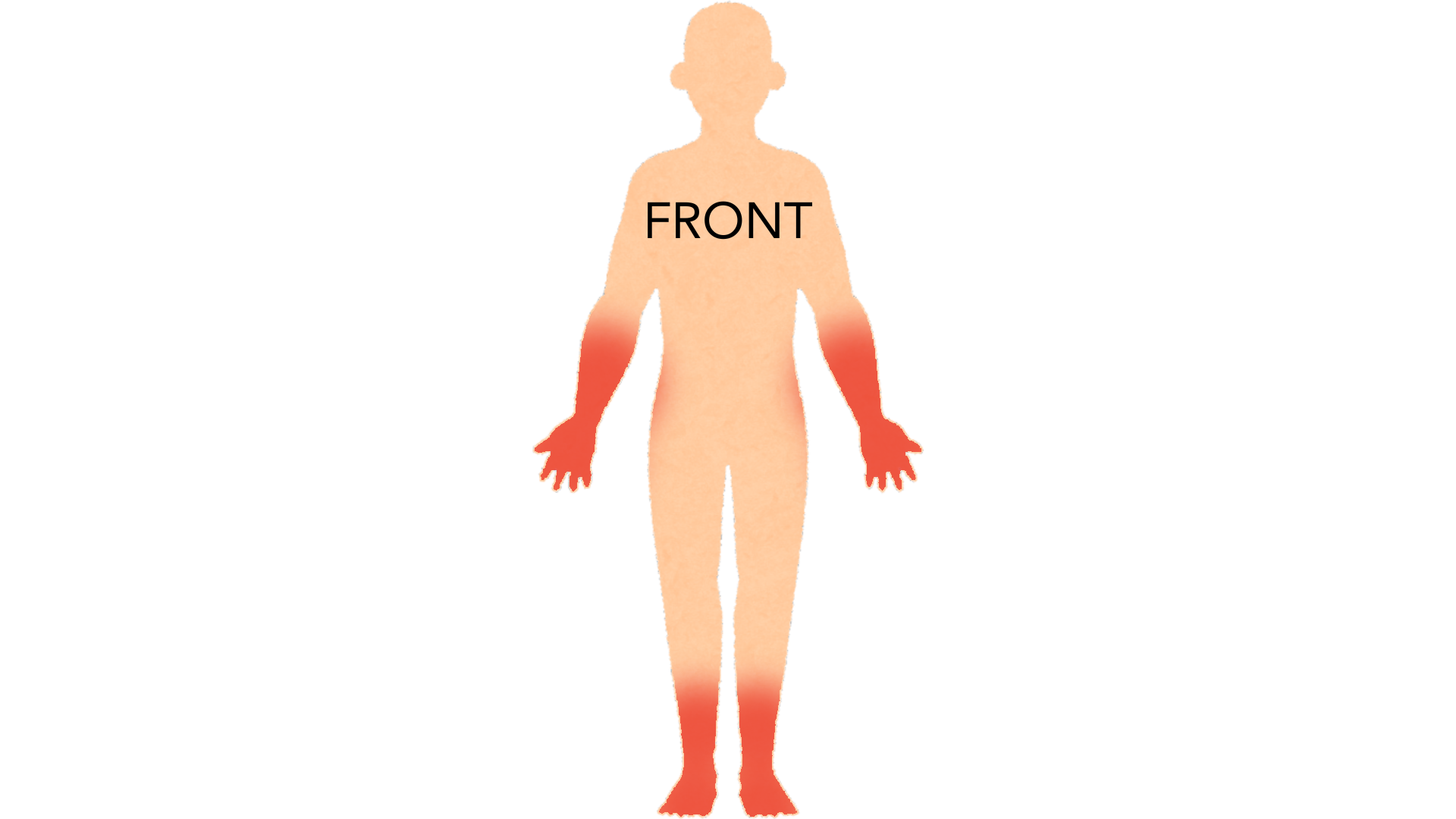
The backs of the hands and the forearms come next. On deeper skin tones the pattern is different: it appears more often on the legs and feet, and inside old burn scars, ulcers and wounds that have never healed, rather than on sun-exposed skin.
Basal Cell Carcinoma
Almost never
Spread beyond the original site is very rare. The damage it does is local.
Squamous Cell Carcinoma
Uncommon, but possible
The risk is higher on the lip and ear, in large or deep tumours, and in people whose immune systems are suppressed. This is why an SCC is usually treated sooner.
Basal Cell Carcinoma
Nothing specific
Nothing reliably comes first. It starts as the bump itself.
Squamous Cell Carcinoma
Actinic keratosis
A small rough, scaly, sandpapery patch on sun-damaged skin. A small fraction of these turn into SCC. That is what your dermatologist is freezing off when they treat rough spots on your face or scalp.
Basal Cell Carcinoma
Cut it out
Most are removed by a simple excision under local anaesthetic. On the face, Mohs surgery is common. The surgeon takes thin layers and checks each one under the microscope until no cancer is left, which spares the most healthy skin.
Squamous Cell Carcinoma
Cut it out, and check further if needed
The same approach, with a wider margin. For higher-risk tumours a doctor may also examine nearby lymph nodes or order imaging.

A small sample taken under local anesthetic, usually a shave or a punch, and read by a pathologist. It does two things: it confirms this is a basal cell carcinoma, and it names the subtype. The subtype is what decides which treatment is right, so this comes before the treatment conversation rather than after it.

The tumor is removed in stages and the edges are examined under a microscope during the same appointment, so complete removal is confirmed before the wound is closed. It has the highest cure rate and spares the most normal tissue, which is why it is used on the face and for aggressive subtypes. It takes several hours, and it is not needed for every tumor.

The tumor is cut out with a margin of normal skin around it and the wound is stitched, with the margins checked by a pathologist afterwards. It is the standard treatment for most tumors away from the face, and it cures the large majority in one visit. If the report comes back with the tumor at the edge, more treatment is needed.
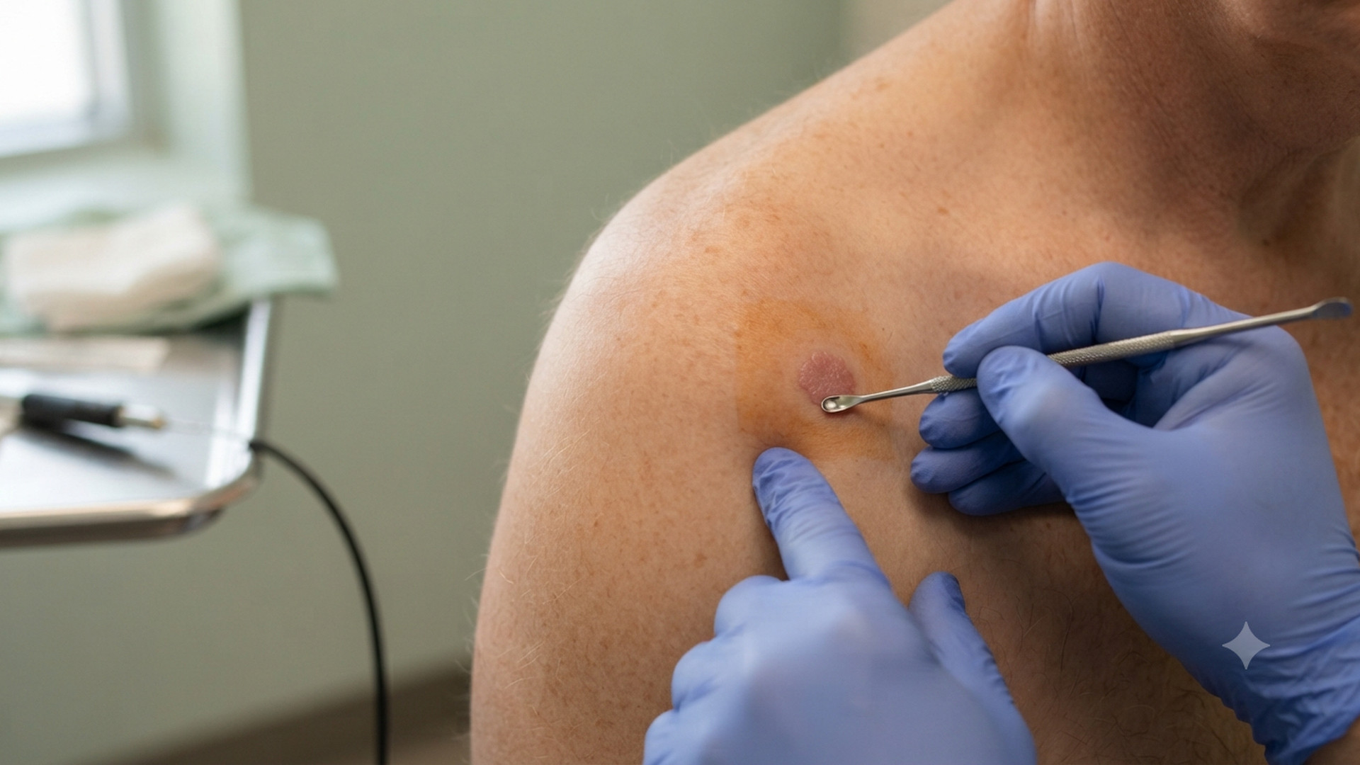
The tumor is scraped away and the base burned, repeated a few times in one visit. It suits small, thin, superficial tumors on the trunk and limbs and is quick and cheap. Nothing is checked under a microscope afterwards, so it is not used on the face or for aggressive subtypes, and it leaves a permanent pale round mark.

A light-sensitizing cream is left on the tumor and then activated with a lamp, usually in two sessions. It is for thin superficial tumors only, confirmed by biopsy, and it has the advantage of treating the sun-damaged skin around them at the same time. It stings during the light, and the cure rate is lower than surgery.
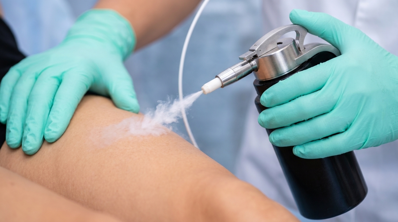
Freezing is quick and occasionally used for small superficial tumors when other options do not suit. Nothing is checked under a microscope, so an incompletely treated tumor can carry on growing under a healed surface. It also leaves a permanent pale patch, which is much more noticeable on deeper skin tones.
Nothing else on this page happens without it. Even an experienced dermatologist with a dermatoscope cannot settle this by looking, and the report says far more than yes or no — how deep it goes, how aggressive the cells look, whether it is tracking along a nerve. Those details choose the treatment. It takes a few minutes under local anesthetic and leaves a small wound. A biopsy does not make cancer spread.
The tumor is removed a layer at a time and each layer is checked under the microscope during the same appointment, so the surgeon stops as soon as the edges are clear. It gives the highest cure rate and takes the least normal skin, which is why it is used on the face, ears, lips, hands, feet and genitals, and for large, recurrent or aggressive tumors. It is a long appointment rather than a difficult one.
The standard removal: the tumor is cut out with a margin of normal skin around it and the wound is stitched closed, usually in one visit under local anesthetic. It cures the large majority of straightforward tumors on the trunk, arms and legs. The margins are checked afterwards rather than during, so occasionally a second procedure is needed to clear an edge.
The tumor is scraped out with a small loop and the base is sealed with an electric current, repeated a few times in one sitting. It is quick and effective for small, thin, low-risk tumors on the trunk and limbs. It is not used on the face, on large or recurrent tumors, or anywhere depth matters, because nothing is sent for margin checking and it leaves a round pale scar.
A light-activated cream treatment for the earliest forms only — actinic keratoses and some cases of carcinoma in situ. It treats a whole patch of sun-damaged skin at once and heals with a good cosmetic result. It has no role in an invasive tumor, and using it on one delays proper removal.
A quick freeze in clinic, used for actinic keratoses and occasionally for a small patch of carcinoma in situ. It is a treatment for the step before this cancer, not for the cancer itself. Freezing an invasive tumor leaves cells behind at depth and hides what is happening under a healing crust.
Basal Cell Carcinoma
Very high
Squamous Cell Carcinoma
Very high
Basal Cell Carcinoma
Squamous Cell Carcinoma

The flat, scaly, pink kind of basal cell carcinoma on the trunk is regularly treated as eczema for months. Eczema itches, comes and goes, appears in more than one place, and settles with a steroid cream. This sits in one place, does not itch much, has a fine shiny thread-like border, and keeps coming back exactly where it was. A single stubborn patch that will not respond deserves a second look.
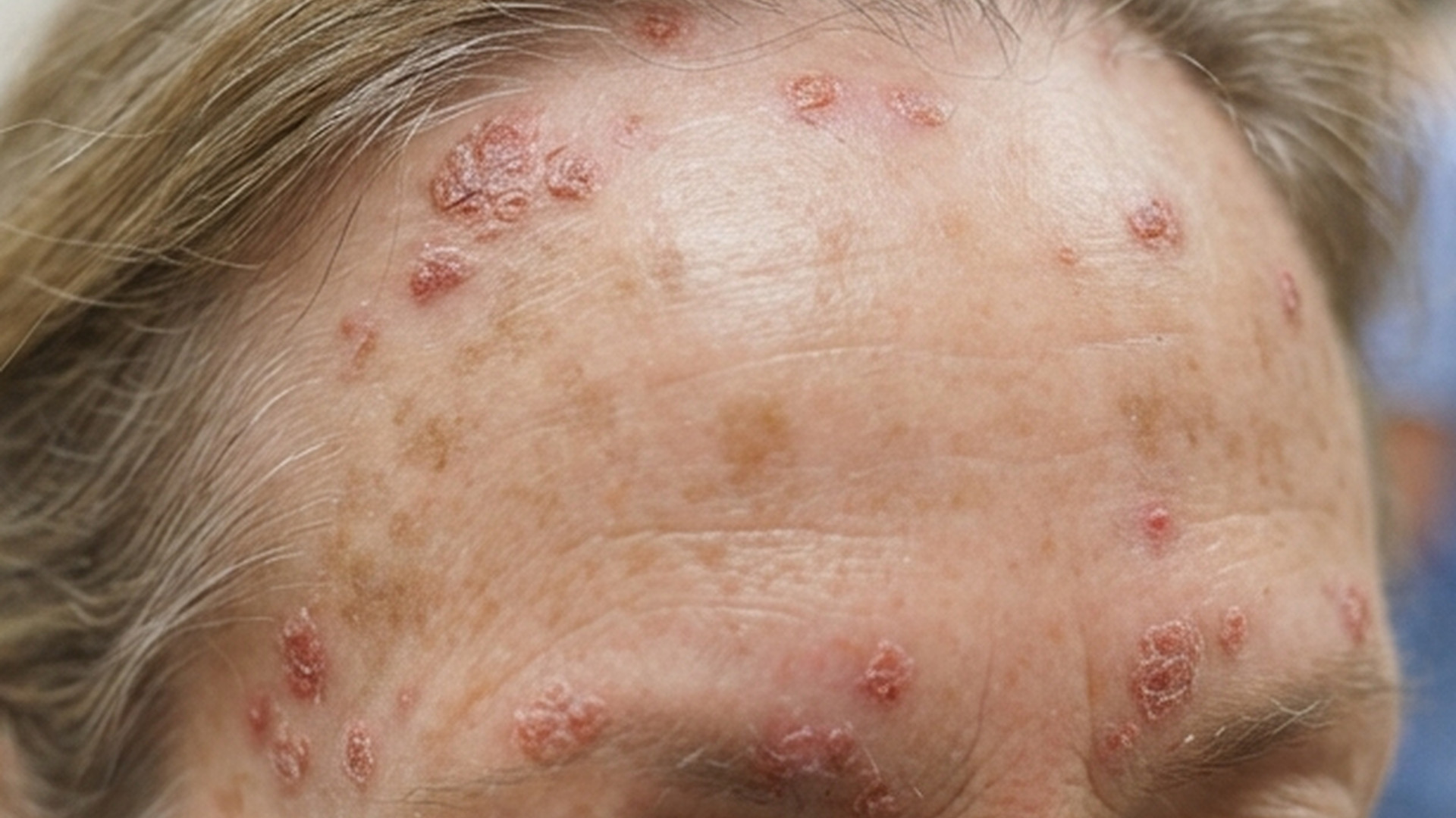
These are the step immediately before this cancer, so they sit on the same sun-damaged skin and feel like the same sandpaper. An actinic keratosis is flat or barely raised, comes in numbers rather than singly, and flakes off and returns. A squamous cell carcinoma is the one that thickens into a firm lump, becomes tender, bleeds, and stays. Any single rough patch that starts to grow up rather than sit flat is the one to have looked at.
Both are common sun-driven cancers, both turn up on the face and ears, and both can present as a sore that will not heal. A basal cell carcinoma tends to look pearly or translucent, with a rolled shiny edge and fine threadlike vessels running across it, and it grows slowly over years. A squamous cell carcinoma is rougher, scalier, more often tender, and moves over weeks to months. Only the biopsy separates them reliably, and both are treated by removal.
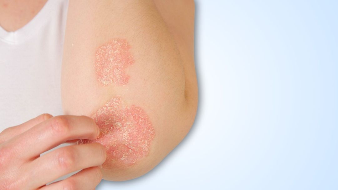
This is the mix-up that costs people the most time. Squamous cell carcinoma in situ on the lower leg is a persistent scaly red or darker patch, and it is regularly treated as psoriasis or eczema for months. Psoriasis is usually symmetrical, turns up on both shins, knees or elbows at once, and improves with the creams given for it. A single patch, on one leg only, that has never responded to those creams should be biopsied rather than treated again.

At the bottom of the outer layer of skin sits a single row of basal cells. They divide at a steady pace to replace the cells shed from the surface above them. Control genes inside each one hold that pace in check, and their DNA is intact.

Ultraviolet light from the sun or a tanning bed damages the DNA inside the basal cells at the bottom of the outer skin layer. Almost all of that damage is repaired. Over decades, a small amount is not, and it builds up in the skin that gets the most exposure.

Enough damage collects in one cell to break a control gene, most often PTCH1, that normally keeps a growth signal switched off. With the switch stuck on, that cell divides without stopping. The bump this makes grows over months and years, not weeks, which is why it is so easy to dismiss.

The tumor works sideways and downward through whatever tissue is next to it. It almost never enters the bloodstream, so it rarely reaches other organs, but on the face it can eventually reach cartilage, muscle, nerve and bone. Nearly all of the harm from this cancer comes from this step, and this step is a function of time.

The skin around the tumor has taken the same lifetime of ultraviolet exposure, which dermatologists call field damage. That is why one basal cell carcinoma predicts more, and why a large proportion of people develop another within five years. Everything on the lists below is either shortening step three or reducing this step.
The outer layer of skin is a stack of flat cells that divide, move up and are shed. Every new copy is checked, and a gene called p53 retires any cell whose DNA is too damaged to fix. The DNA inside those cells is intact, and a firm boundary line underneath holds the whole layer above the deeper skin.

Ultraviolet light, mostly UVB from the sun and from tanning beds, reaches the outer layer of the skin and damages the DNA inside the flat cells there. This happens on ordinary days, not only on sunny holidays, and it adds up over decades.

The body repairs most of that damage, and cells too damaged to repair are supposed to die off. Over years some of the damage lands on the genes that run repair and shutdown, p53 most often, and once those controls are broken the damaged cells stay alive.

The surviving cells divide. First they build up as a rough precancerous patch, an actinic keratosis. Then they can fill the whole thickness of the top layer of skin, which is carcinoma in situ. Everything so far is still held above the boundary line.

The cells break through the boundary into the deeper layer, and the tumor now has access to blood vessels and lymph channels. It grows down into fat, cartilage, nerve or bone, and a small share reach a lymph node. Almost everything on the lists below is pushing on this step, and how early it is removed decides how it goes.
Common questions
Neither of these is melanoma, which is the rarer and more dangerous type of skin cancer.